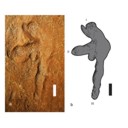
3D models of Miocene vertebrates from Tavers
3D model of Burmesescorpiops wunpawng
3D models of early strepsirrhine primate teeth from North Africa
3D GM dataset of bird skeletal variation
Skeletal embryonic development in the catshark
Bony connexions of the petrosal bone of extant hippos
bony labyrinth (11) , inner ear (10) , Eocene (8) , South America (8) , Paleobiogeography (7) , skull (7) , phylogeny (6)
Lionel Hautier (23) , Maëva Judith Orliac (21) , Laurent Marivaux (16) , Rodolphe Tabuce (14) , Bastien Mennecart (13) , Pierre-Olivier Antoine (12) , Renaud Lebrun (11)
MorphoMuseuM, also referred to as M3, is a peer reviewed, online data journal that publishes 3D models of vertebrates, including models of type specimens, anatomy atlases, reconstruction of deformed or damaged specimens, and 3D datasets (see https://doi.org/10.1017/scs.2017.14 for details).
M3 comes along with a free software, MorphoDig, which contains a set of tools for editing, positioning, deforming, labelling, measuring and rendering sets of 3D surfaces.
All 3D data presented on this website are licensed under a Creative Commons Attribution-NonCommercial 4.0. International License.
|
|
3D models related to the publication: Comparative endocranial traits in the crocodylians Leidyosuchus canadensis and Stangerochampsa mccabei from the upper Cretaceous of Alberta, Canada.Garance Donzé
Published online: 14/01/2026 |
|
|
M3#1830Skull, endocast, paratympanic sinuses, jugal and maxillary neurovascular canals, alveoli Type: "3D_surfaces"doi: 10.18563/m3.sf.1830 state:in_press |
Download 3D surface file |
Stangerochampsa mccabei TMP1986.61.1 View specimen
|
|
M3#1831Skull, endocast, paratympanic sinuses, jugal and maxillary neurovascular canals, alveoli Type: "3D_surfaces"doi: 10.18563/m3.sf.1831 state:in_press |
Download 3D surface file |
Alligator mississipiensis OUVC 9761 View specimen
|
|
M3#1833Skull, endocast, paratympanic sinuses, jugal and maxillary neurovascular canals, alveolies Type: "3D_surfaces"doi: 10.18563/m3.sf.1833 state:in_press |
Download 3D surface file |
Alligator sinensis NHMW-Zoo-HS-37966 View specimen
|
|
M3#1835Skull, endocast, paratympanic sinus, jugal and maxillary neurovascular canals, alveoli Type: "3D_surfaces"doi: 10.18563/m3.sf.1835 state:in_press |
Download 3D surface file |
Crocodylus niloticus MHNL 50001399 View specimen
|
|
M3#1834Skull, endocast, paratympanic sinuses, jugal and maxillary neurovascular canals, alveolies Type: "3D_surfaces"doi: 10.18563/m3.sf.1834 state:in_press |
Download 3D surface file |
Diplocynodon ratelii LA86 View specimen
|
|
M3#1832Skull, endocast, paratympanic sinus, jugal and maxillary neurovascular canals, alveolies Type: "3D_surfaces"doi: 10.18563/m3.sf.1832 state:in_press |
Download 3D surface file |
This contribution contains the 3D models described and figured in: Phylogenetic signal in anteater snout morphology: implications for interpreting rare vermilinguan fossils. Palaeobiodiversity and Palaeoenvironments.
Indet indet VPPLT 977 View specimen
|
|
M3#17933D surface models of the cranium, nasal bone and cranial canals Type: "3D_surfaces"doi: 10.18563/m3.sf.1793 state:in_press |
Download 3D surface file |
Cyclopes didactylus M 1525 View specimen
|
|
M3#17943D models of the cranium and internal cranial canals Type: "3D_surfaces"doi: 10.18563/m3.sf.1794 state:in_press |
Download 3D surface file |
Cyclopes didactylus M 1571 View specimen
|
|
M3#17953D surface models of the cranium, nasal bone and cranial canals Type: "3D_surfaces"doi: 10.18563/m3.sf.1795 state:in_press |
Download 3D surface file |
Myrmecophaga tridactyla M 3023 View specimen
|
|
M3#17963D surface models of the cranium, nasal bone and cranial canals Type: "3D_surfaces"doi: 10.18563/m3.sf.1796 state:in_press |
Download 3D surface file |
Tamandua tetradactyla NHMUK 3.7.7.135 View specimen
|
|
M3#17973D models of the cranium and internal cranial canals Type: "3D_surfaces"doi: 10.18563/m3.sf.1797 state:in_press |
Download 3D surface file |
Tamandua tetradactyla NHMUK 4.7.4.90 View specimen
|
|
M3#17983D surface models of the cranium, nasal bone and cranial canals Type: "3D_surfaces"doi: 10.18563/m3.sf.1798 state:in_press |
Download 3D surface file |
Tamandua tetradactyla UM 788N View specimen
|
|
M3#17993D models of the cranium and internal cranial canals Type: "3D_surfaces"doi: 10.18563/m3.sf.1799 state:in_press |
Download 3D surface file |
Cyclopes didactylus MVZ 121210 View specimen
|
|
M3#18003D models of the cranium and internal cranial canals Type: "3D_surfaces"doi: 10.18563/m3.sf.1800 state:in_press |
Download 3D surface file |
Myrmecophaga tridactyla MVZ 112943 View specimen
|
|
M3#18013D models of the cranium and internal cranial canals Type: "3D_surfaces"doi: 10.18563/m3.sf.1801 state:in_press |
Download 3D surface file |
Myrmecophaga tridactyla MVZ 185238 View specimen
|
|
M3#18023D models of the cranium and internal cranial canals Type: "3D_surfaces"doi: 10.18563/m3.sf.1802 state:in_press |
Download 3D surface file |
|
|
3D Printing an Explodable Dog Skull for Veterinary EducationWilliam C. Hooker
Published online: 17/12/2025 |
|
|
M3#1858PLYs of the segmented cranial bones with pre-fabricated magnetic casings and shelves for assembly following 3D printing Type: "3D_surfaces"doi: 10.18563/m3.sf.1858 state:published |
Download 3D surface file |
|
|
M3#1859PLYs of the segmented cranial bones of the "BOTTOM" cranial component. Downloadable for additional learning opportunities for students Type: "3D_surfaces"doi: 10.18563/m3.sf.1859 state:published |
Download 3D surface file |
|
|
3D models related to the publication: Révision des données sédimentologiques et biostratigraphiques des gisements à vertébrés des sables de l’Orléanais, à Beaugency, Tavers et Le Bardon (Miocène Moyen ; Loiret, France)Adrien de Perthuis
Published online: 31/10/2025 |
|
|
M3#1837Left upper M3 Type: "3D_surfaces"doi: 10.18563/m3.sf.1837 state:published |
Download 3D surface file |
Megamphicyon giganteus ULB-TAV-13 View specimen
|
|
M3#1531Left first lower molar Type: "3D_surfaces"doi: 10.18563/m3.sf.1531 state:published |
Download 3D surface file |
Hispanotherium matritense ULB-TAV-17 View specimen
|
|
M3#1532Left first lower molar Type: "3D_surfaces"doi: 10.18563/m3.sf.1532 state:published |
Download 3D surface file |
Plesiaceratherium lumiarense ULB-TAV-18 View specimen
|
|
M3#1533Left third upper molar Type: "3D_surfaces"doi: 10.18563/m3.sf.1533 state:published |
Download 3D surface file |
Chelydropsis aff. sansaniensis ULB-TAV-23 View specimen
|
|
M3#1535Cast of a skull Type: "3D_surfaces"doi: 10.18563/m3.sf.1535 state:published |
Download 3D surface file |
Ronzotherium romani ULB-TAV-4 View specimen
|
|
M3#1556Right fourth upper premolar Type: "3D_surfaces"doi: 10.18563/m3.sf.1556 state:published |
Download 3D surface file |
Prodeinotherium bavaricum ULB-TAV-24 View specimen
|
|
M3#1557left hemimandibule Type: "3D_surfaces"doi: 10.18563/m3.sf.1557 state:published |
Download 3D surface file |
In this contribution a third new species of the rare genus Burmesescorpiops Lourenço, 2016 is described. The discovery of this new element belonging to the family Palaeoeuscorpiidae Lourenço, 2003 and to the subfamily Archaeoscorpiopinae Lourenço, 2015 brings further elements to support the validity of the genus Burmesescorpiops. This generic group remains however, poorly speciose. This is the latest discovery of Burmesescorpiops wunpawng, the name is derived from the Kachin Hilltribe peoples who are indigenous to the area. The data provided here is a 3D surface.
Burmesescorpiops wunpawng ps-gyi-01-25 View specimen
|
|
M3#18463d Surface Volume Type: "3D_surfaces"doi: 10.18563/m3.sf.1846 state:published |
Download 3D surface file |
This contribution contains the 3D models described and figured in the following publication:Skull and Inner Ear Morphometrics in Sheep and Goats: Species and Breed Differentiation with Bioarchaeological Applications (Hemelsdael et al. submitted). The models include the external surface of a complete skull and inner ear of both a sheep (Ovis aries) and a goat (Capra hircus), generated from micro-CT scans. In the associated paper, we used 3D geometric morphometric data to assess inter and intra (i.e. between breeds) discrimination based on complete skulls, skull fragments and the semi-circular canals of the inner ear.
Capra hircus Amp_1 View specimen
|
|
M3#1806Skull of the goat Amp_1 Type: "3D_surfaces"doi: 10.18563/m3.sf.1806 state:published |
Download 3D surface file |
|
|
M3#1807Inner ear of the goat Amp_1 Type: "3D_surfaces"doi: 10.18563/m3.sf.1807 state:published |
Download 3D surface file |
Ovis aries UM_RR_2331 View specimen
|
|
M3#1808Skull of the sheep UM_RR_2331 Type: "3D_surfaces"doi: 10.18563/m3.sf.1808 state:published |
Download 3D surface file |
|
|
M3#1809Inner ear of the sheep UM_RR_2331 Type: "3D_surfaces"doi: 10.18563/m3.sf.1809 state:published |
Download 3D surface file |