
Pterosaurs from Coahuila
Abstract book of the 18th Conference of the EAVP
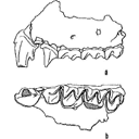
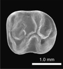
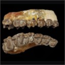
Two enigmatic rodents from Lavergne (MP 16), Quercy Phosphorites
Les sélaciens du Miocène de la région de Montpellier
Muridae du Pliocène supérieur d'Espagne et du midi de la France.
Les Chiroptères du Miocène inférieur de Bouzigues. 1- Etude systématique.
Eocene (57) , Quercy Phosphorites (38) , Systematics (32) , Rodents (29) , Mammalia (27) , Rodentia (25) , Miocene (24)

|
Les sélaciens du Miocène de la région de MontpellierHenri Cappetta
Published online: 15/12/1970 |
|

|
Muridae (Rodentia) du Pliocène supérieur d'Espagne et du midi de la France.Jacques MichauxPublished online: 20/09/1969Keywords: Anthracomys meini; Castillomys crusafonti; Pliocene; Rodents; Valerymys ellenbergeri https://doi.org/10.18563/pv.3.1.1-25 Abstract The murid fauna of the terminal Pliocene of southwest Europe is rich in at least eight genera and ten species. With the species belonging to the genera Apodemus, Rhagapodemus, and Stephanomys not being studied here, the study of the other murids resulted for one thing in the description of three new genera and three new species: Castillomys crusafonti n. g., n. sp., Occitanomys brailloni n. g., n. sp., Anthracomys meini n. sp., Valerymys ellenbergerí (THALER) n. g., and for another thing in the recognition of a form hitherto unknown in this region, Micromys praeminutus KRETZOI. Systematic study has shown that certain species of the terminal Pliocene fauna had their ancestors in the Turolian fauna presently known in Spain. The evolutionary lineages thereby recognized have been studied more in detail and a list of the evolutionary tendencies of the dendal characters has been given. A chart of the probable phyletic relationships between the different murids of the Pliocene faunas of southwest Europe (With the genus Rhagapodemus and Apodemus dominans being excluded) is given in conclusion of this work. PV article infos Published in Vol. 03, Fasc. 1 (1969) |
|
|
Les Chiroptères du Miocène inférieur de Bouzigues. 1- Etude systématique.Bernard SigéPublished online: 17/04/1968Keywords: bats https://doi.org/10.18563/pv.1.3.65-133 Abstract In recent years, the techniques of chemical preparing have permitted a rich paleontologic material to be obtained from the phosporitic sediment of Bouzigues (Hérault, France). The fauna of this locality is comprised of quite varied microvertebrates, amphibians, reptiles, birds, mammals. Twenty five species of the latter, belonging to seven orders, are today known from the site. Among them, the numerous rodents have allowed L. Thaler to chronologically situate this fauna in the Zone of Laugnac (<< late Aquitanian ›> of some authors). PV article infos Published in Vol. 01, Fasc. 3 (1968) |
|

|
Contribution à l'étude des genres Gliravus et Microparamys (Rodentia) de l'Eocène d'Europe.Jean-Louis HartenbergerPublished online: 15/03/1971Keywords: Eocene; Gliravus; Microparamys; Rodentia https://doi.org/10.18563/pv.4.4.97-135 Abstract Based on material found in about 15 localities the relationships of the genera Microparamys and Glirarus have been studied. One new genus, two subgenera and three species [Microparamys (Sparnacomys) chandoni n. subgen. and n. sp., Microparamys (Pantrogna) russelli n. subgen., Eoglirarus wildi n. gen. and n. sp., Gliravus meridionalis n. sp.] as well as the publication PV article infos Published in Vol. 04, Fasc. 4 (1971) |
|

|
Révision des Chiroptères Lutériens de Messel (Hesse, Allemagne).Donald E. Russell and Bernard SigéPublished online: 04/04/1970Keywords: Chiroptera; Lutetian; Messel https://doi.org/10.18563/pv.3.4.83-182 Abstract The revision of the Lutetian chiropterans from Messel, first described by Revilliod in 1917, is based on the anatomy of the teeth and the skeleton. A figuration or refiguration of thematerial utilized accompanies the new description, which goes beyond that of the original monograph. PV article infos Published in Vol. 03, Fasc. 4 (1970) |
|

|
Arvicolinae (Rodentia) du Pliocène terminal et du quaternaire ancien de France et d'Espagne.Jacques MichauxPublished online: 30/10/1971Keywords: Arvicolinae; France; Pleistocene; Pliocene; Spain https://doi.org/10.18563/pv.4.5.137-214 Abstract Two steps can be distinguished in the history of the first invasion of western and south western Europe by the arvicolines. The first step corresponds to the installation of these rodents with the immigration of Promimonys inxuliferus Kowalski, then of Mimomys stehlini Kormos and of Mimomys gracilis (Kretzoi). The second is characterized by the establishment of a geographic differentiation in the arvicoline fauna between the south of France and Spain, from where are described new species of Mimomys (Mimamys cappettai, Mimomys septimanus, Mimonys medasensis), and the rest of France, where are found only elements already known from central Europe or England (Mimomys polonicus Kowalski, Mimomys pliocaenicus F. Major, Mimomys reidi Hinton, or forms very close to the latter). This geographic differentiation, which is very certainly the consequence of the division of Europe into distinct climatic provinces, one of them being the southern province comprising at least Spain and southern France, could result from a cladogenetic evolution of Mimomys stehlini and Mimomys gracilis after their immigration. The present work is also a contribution to the search for correlations between the diverse micromammal localities of the latest Pliocene (or early Villafranchian) and of the early Quaternary of Europe. PV article infos Published in Vol. 04, Fasc. 5 (1971) |
|

|
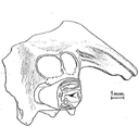
Osteology of Prolagus sardus, a Quaternary Ochotonid (Mammalia, Lagomorpha).Mary R. DawsonPublished online: 21/06/1969Keywords: Lagomorpha; Ochotonidae; Prolagus https://doi.org/10.18563/pv.2.4.157-190 Abstract Prolagus sardus is the last representative of the diverse lineages of European endemic ochotonids. It is also the most abundant in the collections. The previous studies made of this species have established rather well its dental morphology, its phylogenetic position, its geographic and temporal distribution, and its intraspecific individual variation. On the other hand, no osteologic study has fully utilized the superb material from Corsica and Sardinia collected by Forsyth Major. PV article infos Published in Vol. 02, Fasc. 4 (1969) |
|
|
A new and primitive species of Protophiomys (Rodentia, Hystricognathi) from the late middle Eocene of Djebel el Kébar, Central TunisiaLaurent Marivaux
Published online: 02/06/2014 |
|

|
Reflections on some Russian eotheriodonts (Reptilia, Synapsida, Therapsida)Denise Sigogneau-Russell and P. K. TchudinovPublished online: 28/02/1972Keywords: Reptilia; Russia; Synapsida; Therapsida https://doi.org/10.18563/pv.5.3.79-109 Abstract As a result of the enrichment of eotheriodont material by one of us (P.K.T.), these specimens (essentially Biarmosuchur and Eotitanosuchur) are reexamined and refigured. A reevaluation of their particularities supports the distinction of two families, for which new diagnoses are proposed. This leads us to discuss the affinities of these families, with respect to the sphenacodonts on one hand, and to the South African primitive theriodonts on the other (gorgonopsids and ictidorhinids). This study contains inherent paleogeographic consequences which are considered in conclusion. PV article infos Published in Vol. 05, Fasc. 3 (1972) |
|

|
Les gisements de Robiac (Eocène supérieur) et leurs faunes de Mammifères.Jean SudrePublished online: 05/04/1969Keywords: Fauna; Late Eocene; Mammalia; Robiac https://doi.org/10.18563/pv.2.3.95-156 Abstract Designated the type-locality of a late Eocene paleomammal zone, Robiac has recently been the object of important excavations. The first results of the new collecting, as well as a revision of the material in old collections, are given in this work. PV article infos Published in Vol. 02, Fasc. 3 (1969) |
|

|
Rongeurs nouveaux de l'Oligocène Moyen d'Espagne.Louis ThalerPublished online: 15/09/1969Keywords: Cricetidae; Oligocene; Pseudocricetodon; Rodents; Theridomys https://doi.org/10.18563/pv.2.5.191-207 Abstract Description of four new rodents from a recently discovered locality at Montalban. Theridomys crusafonti nov. sp. is considered as the ancestry of T. Iembronicus. Theridomys varian: nov. sp. includes «Theridomys» morphotypes and «Blainvilllimys» morphotypes; it could be ancestral to B. blainvillei. Pseudoltinomys nanus nov. sp. represents a new lineage paralleling in evolution that of P. gaillardi (which is equally found at Montalban). Pseudocricetodon montalbanensis nov. gen., nov. sp. designates a lineage of very small Cricetidae accompanying Eucricetodon. With these well defined new species and six others present in the locality, Montalban appears as the best faunal reference point within the biochronologic zone of La Sauvetat. PV article infos Published in Vol. 02, Fasc. 5 (1969) |
|

|
Les Pseudosciuridae (Mammalia, Rodentia) de l'Eocène moyen de Bouxwiller, Egerkingen et Lissieu.Jean-Louis HartenbergerPublished online: 30/10/1969Keywords: Bouxwiller; cranium; Egerkingen; Middle Eocene; Rodents https://doi.org/10.18563/pv.3.2.27-64 Abstract The description of new material from three classic middle Eocene localities of western Europe permits the addition of details to the systematics of primitive Pseudosciurids. The cranial anatomy of Protadelomys cartieri (STEHLIN and SCHAUB) from Egerkingen is described here and compared to that of the Adelomyines from the late Eocene, until now the only ones known. The morphologic and biometric study of the dentition of P. cartieri compared to that of P. alsaticus n. sp. from Bouxwiller and P. Iugdunensis n. sp. from Lissieu, forms respectively older and younger than P. cartieri, permits the evolutionary tendencies of the group to be demonstrated and shows that notable differences in age exist between these localities. This ensemble of forms can constitute a valuable guide lineage in the establishment of a fine stratigraphy of the period. Other less well known lineages are present at Egerkingen along with P. cartieri. They can be related to genera that have been noted int he late Eocene. In conclusion, a criticism of recent zonation proposals, made by divers authors, completes this article. PV article infos Published in Vol. 03, Fasc. 2 (1969) |
|

|
Les traces de pas d'amphibiens, de dinosaures et autres reptiles du Mesozoïque Français : inventaire et interprétations.Georges Gand, Georges Demathieu and Christian MontenatPublished online: 15/12/2007Keywords: Footprints; France; Inventory; Mesozoic; palaeontology; palaeovenvironments; Stratigraphy https://doi.org/10.18563/pv.35.1-4.1-149 Abstract Since the 19th century, thousands of footprints were observed in the geological series of the French Mesozoic. All are located in the Triassic and Jurassic. After a promising beginning, in France, it is only a few papers which will be published in the first half 20th century, unlike the USA and of others countries of Western Europe. One ought to wait about 1950 for a revival and now they are nearly 200 papers which were devoted to the ichnofossils. The literature abundance and the renewed interest of the naturalists for the palichnologic studies decided to us to write a synthesis work. This one begins with a stratigraphic inventory in which, localisation, age and paleontological contents of about 180 fossiliferous sites are specified. After having pointed out the followed methods, the footprints paleontological interpretation is then approached in detail and the results obtained are replaced in stratigraphy to deduce the fauna evolution during the Mesozoic. So, it appears that Ichnologic data, more varied and rich in the Triassic and Liassic than those relating to the bones, very rare for the considered periods, are very informative. The middle Triassic (Anisian-Ladinian), thus reveals Cotylosauria, Lepidosauria, Crurotarsi with Rauisuchia, Ornithosuchidae, Crocodylia and Dinosauromorpha more the "Prodinosauria": Dinosamiforme whose skeletons are known in Argentina but only in Ladinian. The rather fast domination of Dinosaurs during Norian is also as well shown. The almost exclusive presence of their footprints, up to fifty cm long, in the Lower Hettangian indicates their supremacy in the environments. Footprints characterise not very deep life places located between inter-supratidal limits and often out of water. Sedimentologic and Palaeontologic studies showed that they were great coastal spaces during Middle Triassic, flood-plain with sebkhas while Upper Triassic, and a large !!coastal marsh!! in Grands-Causses during Liassic in which, mainly, fine stromatolithic layers were deposited. During the same periad, bay beaches spread in Vendée. During the Middle Jurassic, they are also brackish to lacustrine environments and recifallagoons in- the Upper Jurassic. Numerous measurements of the footprints and trackways directions showed that the animaIs moved there in weil defined directions, for long periods. They seem due to the palaeotopography of the life environments relatively stable. Also, the discovery of vegetal radicular networks and small footprints far away from the continental borderlands has suggested that the animals continuously lived in these palaeoenvironnements, belonging to large ecosystems, where the sedimentation rate was weak. This explains that thebadies could not fossilize there but only their footprints through the cyanobacterian action in main cases. From the vertical distribution of different ichnospecies, defined with adapted statistical methods, explained in this work, a palichnostratigraphy was established for the Middle Triassic. Although the footprints are also abundant in Hettango-Sinemurian of "Grands-Causses" and the Vendée, it was not possible, up to now, to establish any zonation in this series; Probably because the palichnofauna is too little diversified there, currently reduced to a majority of Theropods II-IV tridactyl traces. PV article infos Published in Vol. 35, Fasc. 1-4 (2007) |
|
|
Les Paramyidae (Rodentia) de l'Eocène inférieur du bassin de Paris.Jacques MichauxPublished online: 15/07/1968Keywords: Ailuraviinae; Eocene; Paramyinae; Rodents https://doi.org/10.18563/pv.1.4.135-193 Abstract The exploitation of new early Eocene localities in the Paris Basin has resulted in the collecting of numerous mammalian remains, among which are about 300 isolated teeth representing the rodents. They belong, for the most part, to the paramyid group. Only the latest level of the early Eocene has yielded rodents belonging to the pseudosciurid group. The paramyids, the object of this study, are represented by at least 5 genera and 10 species; they are distributed among 4 clearly dilferentiated subfamilies : Paramyinae Simpson 1945, Pseudoparamyinae Michaux 1964, Ailuraviínae n. subf., Microparamyinae Wood1962. PV article infos Published in Vol. 01, Fasc. 4 (1968) |
|

|
Contribution à l'étude des Cricétidés oligocènes d'Europe occidentaleMonique Vianey-Liaud
Published online: 20/01/1972 |
|
|
Les Périssodactyles (Mammalia) du gisement Bartonien supérieur de Robiac (Éocène moyen du Gard, Sud de la France)Jean-Albert RemyPublished online: 04/05/2015Keywords: Chasmotherium; new species; Palaeotheriidae; paleoenvironments https://doi.org/10.18563/pv.39.1.e3 Abstract We present here a new updated counting of the perissodactyls of Robiac, the type locality of the MP 16 level of the biochronological scale of paleogene mammals and that of the Robiacian stage of Eocene Land Mammals Ages in Western Europe. PV article infos Published in Vol.39-1 (2015) |
|
|
Fossil snakes from the Palaeocene of São José de Itaboraí, Brazil Part III. Ungaliophiinae, Booids incertae sedis, and Caenophidia. Summary, update and discussion of the snake fauna from the localityJean-Claude Rage
Published online: 16/12/2008 |
|
|
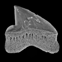
New Squalicorax species (Neoselachii: Lamniformes) from the Lower Maastrichtian of Ganntour phosphate deposit, MoroccoHenri Cappetta
Published online: 05/12/2014 |
|
|
Les Palaeotheridae (Perissodactyla) de la faune de Mammifères de Fons 1 (Eocène supérieur).Jean-Albert RemyPublished online: 15/06/1967Keywords: Anchilophus; Eocene; Pachynolophus; Palaeotheriidae; Perissodactyla https://doi.org/10.18563/pv.1.1.1-46 Abstract The locality of Fons 1, one of the fossiliferous outcrops in the late Eocene limestones of Fons-outre-Gardon (Gard), has yielded varied remains of mammals. The specimens were prepared by dilute acetic acid attack on the rock and by impregnation with an acrylic resin. PV article infos Published in Vol. 01, Fasc. 1 (1967) |
|
|
An evening bat (Chiroptera: Vespertilionidae) from the late Early Eocene of France, with comments on the antiquity of modern batsSuzanne J. Hand
Published online: 01/08/2016 |
|