
Pterosaurs from Coahuila
Abstract book of the 18th Conference of the EAVP
Two enigmatic rodents from Lavergne (MP 16), Quercy Phosphorites
Les sélaciens du Miocène de la région de Montpellier
Muridae du Pliocène supérieur d'Espagne et du midi de la France.
Les Chiroptères du Miocène inférieur de Bouzigues. 1- Etude systématique.
Eocene (57) , Quercy Phosphorites (38) , Systematics (32) , Rodents (29) , Mammalia (27) , Rodentia (25) , Miocene (24)

|
First evidence of an early Miocene marine teleostean fish fauna (otoliths) from la Paillade.(Montpellier,France)Bettina Reichenbacher
Published online: 15/06/1999 |
|

|
Les rongeurs du site Pliocène à Hominidés de Hadar (Ethiope)Maurice SabatierPublished online: 15/02/1982Keywords: Ethiopia; hominids; Muridae; Pliocene https://doi.org/10.18563/pv.12.1.1-56 Abstract The intensive exploration of the Pliocene Hadar Formation, rich in hominid remains, led us to the discovery of several micromammals levels. ln some of them, rodents are very abundant. The stratigraphic repartition of these levels do not cover the whole fossiliferous series of the formation but takes place only in the sedimentary members from Sidi Hakoma and Denen-Dora (rancing from 3.1 - 3.2 MY to 2.8 - 2.9 MY, according to the recent geochronological data). During this gap of time, the species do not show morphological changes, what allowed us to gather, in the same taxa, forms of slighty different ages. PV article infos Published in Vol. 12, Fasc. 1 (1982) |
|
|
Rodent paleocommunities from the Oligocene of Ulantatal (Inner Mongolia, China)Helder Gomes Rodrigues
Published online: 10/06/2014 |
|

|
Les gisements de Robiac (Eocène supérieur) et leurs faunes de Mammifères.Jean SudrePublished online: 05/04/1969Keywords: Fauna; Late Eocene; Mammalia; Robiac https://doi.org/10.18563/pv.2.3.95-156 Abstract Designated the type-locality of a late Eocene paleomammal zone, Robiac has recently been the object of important excavations. The first results of the new collecting, as well as a revision of the material in old collections, are given in this work. PV article infos Published in Vol. 02, Fasc. 3 (1969) |
|

|
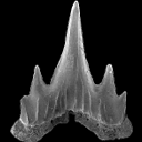
Physogaleus hemmooriensis (Carcharhinidae, Elasmobranchii), a new shark species from the early to middle Miocene of the north sea basin.Thomas Reinecke and Kristiaan HoedemakersPublished online: 15/10/2006Keywords: Carcharhinidae; Early Miocene; Elasmobranchii; Hemmoorian; new species; North Sea Basin; Physogaleus https://doi.org/10.18563/pv.34.e14 Abstract A new carcharhinid shark species, Physogaleus hemmooriensis sp. nov., is described from the Lower Hemmoorian (Behrendorfian, late Burdigalian, early Miocene) of Werder, Lower Saxony, Germany. P. hemmooriensis also occurs in the Edegem and Antwerpen Sands Members of the Berchem Formation, Belgium, and in the Miste Bed, Aalten Member of the Breda Formation, The Netherlands, which have an early to middle Miocene age. In the Western Atlantic region, the taxon is present in the early Miocene Calvert Formation of Delaware, U.S.A, which is largely contemporaneous with the Hemmoorian. PV article infos Published in Vol. 34, Fasc. 1-2 (2006) |
|
|
|
|

|
Study of the Turolian hipparions of the lower Axios valley (Macedonia, Greece). 4. Localities of Dytiko.George D. KoufosPublished online: 15/12/1988Keywords: Equidae; Greece; Hipparion; Lower Axios Valley; Macedonia; Mammalia; Turolian https://doi.org/10.18563/pv.18.4.187-239 Abstract The hipparions from the Dytiko localities of the lower Axios valley (Macedonia, Greece) are studied. The material comes from three localities Dytiko-l, 2, 3 (DTK, DIT, DKO), which are situated near the village of Dytiko, about 60 km northwest to Thessaloniki. Three species have been determined, the medium-sized H. mediterraneum, the small-sized H. matthewi and the very small-sized H. periafricanum. The determined Hipparion species, their morphological characters and their comparison with the other Axios valley material indicate a Late Turolian age for the Dytiko localities. PV article infos Published in Vol. 18, Fasc. 4 (1988) |
|

|
Types dentaires adaptatifs chez les sélaciens actuels et post-paléozoïques.Henri Cappetta
Published online: 01/09/1986 |
|

|
Etude de la Variabilité chez Lophiodon lautricense NouletJean SudrePublished online: 28/02/1971Keywords: Cheek teeth; Eocene; Lophiodon; variability https://doi.org/10.18563/pv.4.3.67-95 Abstract The biometric and morphologie variability of the cheek teeth in the end-of-the-phylum species Lophiodon lautricense Noulet studied in this note, reposes on the observation of about 800 teeth. These were revealed to be little variable in absolute dimensions. The considerable morphologie variability in the upper premolars permitted the problem of the molarization process to be taken up. An hypothesis concerning the order of eruption of the cheek teeth is formulated based on an examination of a large number of milk dentitions. In conclusion, it is suggested that reservations be held on the value of dental characters classically used in systematics for the group under consideration. PV article infos Published in Vol. 04, Fasc. 3 (1971) |
|

|
Les traces de pas d'amphibiens, de dinosaures et autres reptiles du Mesozoïque Français : inventaire et interprétations.Georges Gand, Georges Demathieu and Christian MontenatPublished online: 15/12/2007Keywords: Footprints; France; Inventory; Mesozoic; palaeontology; palaeovenvironments; Stratigraphy https://doi.org/10.18563/pv.35.1-4.1-149 Abstract Since the 19th century, thousands of footprints were observed in the geological series of the French Mesozoic. All are located in the Triassic and Jurassic. After a promising beginning, in France, it is only a few papers which will be published in the first half 20th century, unlike the USA and of others countries of Western Europe. One ought to wait about 1950 for a revival and now they are nearly 200 papers which were devoted to the ichnofossils. The literature abundance and the renewed interest of the naturalists for the palichnologic studies decided to us to write a synthesis work. This one begins with a stratigraphic inventory in which, localisation, age and paleontological contents of about 180 fossiliferous sites are specified. After having pointed out the followed methods, the footprints paleontological interpretation is then approached in detail and the results obtained are replaced in stratigraphy to deduce the fauna evolution during the Mesozoic. So, it appears that Ichnologic data, more varied and rich in the Triassic and Liassic than those relating to the bones, very rare for the considered periods, are very informative. The middle Triassic (Anisian-Ladinian), thus reveals Cotylosauria, Lepidosauria, Crurotarsi with Rauisuchia, Ornithosuchidae, Crocodylia and Dinosauromorpha more the "Prodinosauria": Dinosamiforme whose skeletons are known in Argentina but only in Ladinian. The rather fast domination of Dinosaurs during Norian is also as well shown. The almost exclusive presence of their footprints, up to fifty cm long, in the Lower Hettangian indicates their supremacy in the environments. Footprints characterise not very deep life places located between inter-supratidal limits and often out of water. Sedimentologic and Palaeontologic studies showed that they were great coastal spaces during Middle Triassic, flood-plain with sebkhas while Upper Triassic, and a large !!coastal marsh!! in Grands-Causses during Liassic in which, mainly, fine stromatolithic layers were deposited. During the same periad, bay beaches spread in Vendée. During the Middle Jurassic, they are also brackish to lacustrine environments and recifallagoons in- the Upper Jurassic. Numerous measurements of the footprints and trackways directions showed that the animaIs moved there in weil defined directions, for long periods. They seem due to the palaeotopography of the life environments relatively stable. Also, the discovery of vegetal radicular networks and small footprints far away from the continental borderlands has suggested that the animals continuously lived in these palaeoenvironnements, belonging to large ecosystems, where the sedimentation rate was weak. This explains that thebadies could not fossilize there but only their footprints through the cyanobacterian action in main cases. From the vertical distribution of different ichnospecies, defined with adapted statistical methods, explained in this work, a palichnostratigraphy was established for the Middle Triassic. Although the footprints are also abundant in Hettango-Sinemurian of "Grands-Causses" and the Vendée, it was not possible, up to now, to establish any zonation in this series; Probably because the palichnofauna is too little diversified there, currently reduced to a majority of Theropods II-IV tridactyl traces. PV article infos Published in Vol. 35, Fasc. 1-4 (2007) |
|
|
Critical comments on the genus Propachynolophus Lemoine, 1891 (Mammalia, Perissodactyla, Equoidea)
|
S.I. Data |
|
A new species of hippopotamine (Cetartiodactyla, Hippopotamidae) from the late Miocene Baynunah Formation, Abu Dhabi, United Arab EmiratesJean-Renaud Boisserie
Published online: 07/04/2017 |
S.I. Data |
|
A new species of Propalaeotherium (Palaeotheriidae, Perissodactyla, Mammalia) from the Middle Eocene locality of Aumelas (Hérault, France).Jean-Albert Remy, Gabriel Krasovec
Published online: 24/05/2016 |
S.I. Data |
|
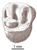
A new and primitive species of Protophiomys (Rodentia, Hystricognathi) from the late middle Eocene of Djebel el Kébar, Central TunisiaLaurent Marivaux
Published online: 02/06/2014 |
|
|
New Late Miocene plecotine bats (Chiroptera, Vespertilionidae: Plecotini) from Gritsev, UkraineValentina V. Rosina, Sergei Kruskop
Published online: 07/03/2019 |
|

|
Contributions à l'étude du gisement Miocène supérieur de Montredon (Hérault). Les grands mammifères. 4 - Les artiodactyles Suidae.Léonard GinsburgPublished online: 15/11/1988Keywords: Artiodactyla; France; Mammalia; Montredon; Upper Miocene https://doi.org/10.18563/pv.18.ext.57-64 Abstract There is only one suid known in the Upper Miocene of Montredon (Hérault): Microstonyx (Limnostonyx nov. subgen.) antiquus (KAUP). It is differenciated from Microstonyx major by the presence of upper and lower canines which are considerably longer and biger. Its presence at Montredon corroborates the palustrine habitat for the species. PV article infos Published in Vol. 18, Ext (1988) |
|
|
New datation of the Tafna Basin (Algeria): A combination between biochronological and magnetostratigraphical dataSalamet Mahboubi
Published online: 11/03/2015 |
|
|
Two new scyliorhinid shark species (Elasmobranchii, Carcharhiniformes, Scyliorhinidae), from the Sülstorf Beds (Chattian, Late Oligocene) of the southeastern North Sea Basin, northern Germany.Thomas ReineckePublished online: 30/04/2014Keywords: Chattian; Elasmobranchii; North Sea Basin; Scyliorhinidae; Scyliorhinus https://doi.org/10.18563/pv.38.1.e1 Abstract Based on isolated teeth two new scyliorhinid shark species, Scyliorhinus biformis nov. sp. and Scyliorhinus suelstorfensis nov. sp., are described from the Sülstorf Beds, early-middle Chattian, of Mecklenburg, northeastern Germany. They form part of a speciose assemblage of necto-benthic sharks and batoids which populated the warm-temperate to subtropical upper shelf sea of the south-eastern North Sea Basin. PV article infos Published in Vol.38-1 (2014) |
|

|
Strange Eocene rodents from SpainPablo Pelaez-Campomanes
Published online: 16/12/1996 |
|

|
Reflections on some Russian eotheriodonts (Reptilia, Synapsida, Therapsida)Denise Sigogneau-Russell and P. K. TchudinovPublished online: 28/02/1972Keywords: Reptilia; Russia; Synapsida; Therapsida https://doi.org/10.18563/pv.5.3.79-109 Abstract As a result of the enrichment of eotheriodont material by one of us (P.K.T.), these specimens (essentially Biarmosuchur and Eotitanosuchur) are reexamined and refigured. A reevaluation of their particularities supports the distinction of two families, for which new diagnoses are proposed. This leads us to discuss the affinities of these families, with respect to the sphenacodonts on one hand, and to the South African primitive theriodonts on the other (gorgonopsids and ictidorhinids). This study contains inherent paleogeographic consequences which are considered in conclusion. PV article infos Published in Vol. 05, Fasc. 3 (1972) |
|