|
Pterosaurs (Pterosauria) from the Cerro del Pueblo Formation (Late Campanian) of Coahuila, Mexico
Published online: 21/11/2025
Keywords:
Azhdarchoidea; Coahuila; Mexico; Pterodactyloidea; Pterosauria
https://doi.org/10.18563/pv.48.2.e1
Abstract
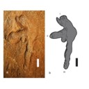
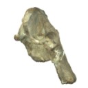
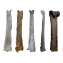
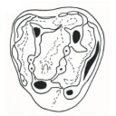
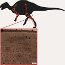
The Late Campanian Cerro del Pueblo Formation, located in southeastern Coahuila, Mexico, has produced a diverse array of vertebrate fossils. However, pterosaur remains from this unit are notably scarce. In this study, we describe new pterosaur material from the formation. The specimens include a fragmentary vertebra identified as belonging to an indeterminate, but derived pterodactyloid, along with the distal condyle of a left metacarpal, referable to an azhdarchoid pterosaur, and a left manus print. While these specimens provide additional evidence of pterosaur presence in the region during the Late Cretaceous, their fragmentary nature limits precise taxonomic and ichnotaxonomic identification. Nevertheless, they highlight the potential for future discoveries that could refine our understanding of the diversity and distribution of pterosaurs in Mexico.
PV article infos
Published in 48-2 (2025)
|
PDF
|
|
Abstract book of the 18th Conference of the European Association of Vertebrate Palaeontologists (EAVP), 5-9 July 2021, Benevento, Italy
Published online: 12/07/2021
Keywords:
2021; Abstracts; Benevento; EAVP
https://doi.org/10.18563/pv.eavp2021
Abstract
Welcome to the 18th conference of the EAVP, the first online meeting of our association. The pandemic emergency made it impossible to organize the in-person meeting in Benevento as we all had hoped. However, we couldn’t miss another EAVP meeting. Therefore, this year we are meeting online, trying to make the experience the closest to the in-person meeting possible, in order to offer the delegates the opportunity to share knowledge, build new networks and reinforce the old ones. We have received 137 communications, with more than 150 delegates from 24 countries. All the abstracts have passed a peer review process and are part of this special volume of Palaeovertebrata, the official journal of the EAVP. This year we are also offering a variety of workshops, roundtables and symposia on different topics. These include the annual “Pride EAVP: An LGBTQ+ Roundtable” and “Women in Palaeontology Roundtable Discussion”, together with the workshops on “Gendered Perspective in Palaeontological Research: from Definition to Action”, “International Palaeontology Education: Virtual Teaching and Real-World Learning”, “Stepping out of Academia: Why, When and How?”, “Introduction to Hypothesis Testing in Statistics”, “The Early-Middle Pleistocene Transition: Marked Mammal Turnover and Ecosystem Dynamic” (included in the early event for the XXI INQUA Congress in Rome 2023, “A Mediterranean Perspective on Quaternary Sciences”). To conclude, we are hosting two symposia on “Palaeoart: Diversity on and behind the Canvas” and “3D fossils, Robotic and Experimental Palaeontology”. We wish you all a happy and productive meeting. And see you in Benevento next year!
PV article infos
Published in Special Volume 1-2021 (2021)
|
PDF
|

|
Mammals of the Eocene locality Toru Ajgyr (Kyrgyzstan)
Published online: 15/12/2006
Keywords:
Eocene; Kyrgyzstan; Mammalia; Olsenia; Palaeoecology; Stratigraphy; taxonomy
https://doi.org/10.18563/pv.34.e12
Abstract
Morphological descriptions are given of Eocene mammals from the locality Toru Ajgyr (NEKyrgyzstan) that were excavated in 1997 and 1998 in a cooperation between the Martin-Luther-University Halle (Germany), the Zoological Institute in St. Petersburg (Russia) and the Seismological Institute in Bishkek (Kyrgyzstan). The species found belong mostly to perissodactyls, as Lophialetes sp., Teleolophus sp. and brontotheres. The primitive ungulate family Olseniidae is represented by a complete foot skeleton of cf. Olsenia sp. In addition, postcranial materials of Gobiatherium mirificum (Dinocerata) and of artiodactyls have been collected and are described herein. Based on mammals, the locality is part of the Asian Land Mammal Age Arshantan and is stratigraphically equivalent with the Bridgerian Land Mammal Age in North America and with the lower and middle Geiseltalian of the European Middle Eocene.
PV article infos
Published in Vol. 34, Fasc. 3-4 (2006)
|
PDF
|
|
Morphological description and identification of an extraordinary new elephant cranium from the early Pliocene of Ileret, Kenya
Published online: 21/10/2021
Keywords:
Elephantidae; Loxodonta adaurora; cranium; early Pliocene; Ileret; Kenya
https://doi.org/10.18563/pv.44.2.e3
Abstract
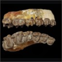
Abstract: Paleontological exploration in the Turkana Basin near Ileret, Kenya yielded the most complete adult elephant cranium (KNM-ER 63642) known from the late Miocene to mid-Pliocene. KNM-ER 63642 derives from the lower Lonyumun Mb. of the Koobi Fora Fm. and dates to the early Pliocene, >4.3 Ma. The cranium is immense in size and preserves most of its structures including left and right M2-3, permitting its comprehensive comparative study and secure taxonomic assignment to Loxodonta adaurora. Features distinctive of the species and exhibited by KNM-ER 63642 include very elongate, divergent tusk alveoli, a short, biconvex cranial roof, anterosuperior angulation of the occipital planum, non-inflated occipital planum and absence of supralateral parietal "bossing," broad, flat premaxillary nasal processes, broad, laterally downturned nasal aperture superior to the level of the orbits, and M3s with wide, subhypsodont plates that are parallel-faced and separated by U-shaped transverse valleys. The M3s also exhibit characteristic L. adaurora traits of greatest width at their bases, rounded cross-sectional shape, thick enamel, abundant cementum, and strong anterior and posterior accessory conules. Of extant taxa, KNM-ER 63642 most closely resembles crania of African elephants. Its inclusion in the Loxodonta clade is tenuous, however, because shared features are either symplesiomorphic or are difficult to test for synapomorphy due to the poor fossil record of crania of late Miocene-early Pliocene elephants. Overall, the cranial morphology of KNM-ER 63642 is unexpectedly advanced for an elephant of its antiquity. Its anteroposterior compression and height are concordant with efficient proal masticatory action, indicating that by the early Pliocene L. adaurora evolved craniodental adaptations in phase with feeding preference for C4 grasses. The advantage of synchrony of morphology and behavior is reflected by the dominance of the species in the greater Turkana Basin during that interval.
PV article infos
Published in 44-2 (2021)
|
PDF
|

|
Contributions à l'étude du gisement Miocène supérieur de Montredon (Hérault). Les grands mammifères. 1 - Les Lagomorphes
Published online: 15/11/1988
Keywords:
Lagomorpha; Montredon; Prolagus; Upper Miocene
https://doi.org/10.18563/pv.18.ext.3-14
Abstract
A sample of 231 isolated teeth of lagomorphs from the upper Miocene of Montredon (southern France), identified as the ochotonid Prolagus crusafonii DOPEZ, 1975, is studied, comparing it with other populations of the same species as well as with its closest species P. oeningensis (KÖNIG, 1825).
The maintenance dining 5 m.y. of a high morphological variability in a group of non-selected features is interpreted as an evolutionary response. This may be related with the heterogeneity and probably too with the regression of the environment.
PV article infos
Published in Vol. 18, Ext (1988)
|
PDF
|
|
Les Chiroptères du Miocène inférieur de Bouzigues. 1- Etude systématique.
Published online: 17/04/1968
Keywords:
bats
https://doi.org/10.18563/pv.1.3.65-133
Abstract
In recent years, the techniques of chemical preparing have permitted a rich paleontologic material to be obtained from the phosporitic sediment of Bouzigues (Hérault, France). The fauna of this locality is comprised of quite varied microvertebrates, amphibians, reptiles, birds, mammals. Twenty five species of the latter, belonging to seven orders, are today known from the site. Among them, the numerous rodents have allowed L. Thaler to chronologically situate this fauna in the Zone of Laugnac (<< late Aquitanian ›> of some authors).
The chiropterans are, with the rodents, the best represented of the locality's mammals. Three families comprise the bat fauna, with nearly complete dominance by one of them (Hippoxideridae) over the two others (Megadermatidae and Vespertílionidae)
Six forms are described, of which three are new species and one a new sub-genus.
Megaderma braillomi n. sp., an animal of rather large size, shows like the Miocene megaderms several evolved dental characters, translating the adaptation of these animals to a partially carnivorous regime. The Bouzigues species seems, however, to represent a particular lineage.
Hipposideros (Brachipposideros n. subgen.) dechaseauxi n. sp. and Hípposideros (Brachipposideros) cf. collongenris Depéret, small sized forms, belong to a group rather well represented in the late Oligocene and early Miocene of Europe, and not distinguished until now within the genus Hipposideros.
Hipposideros (Pseudorhinolophus) bouziguensis n. sp., is the most abundant mammal in the locality and, occuring at the Oligocene-Miocene limit, the last representative known of the subgenus Pseudorhínolophus, common in Europe from the middle Eocene.
However, beyond Neogene and Quaternary times, certain among the numerous living species of Hipposideros are close to Pseudorhinolophus and others to Brachipposíderos. 'This fact would in the future justify a global revision of the genus, on the basis of comparative anatomy of the squeleton and of the teeth.
The bat fauna of Bouzigues is completed by two small Vespertilionidae, rare forms, Myoris sp. I and sp. II.
PV article infos
Published in Vol. 01, Fasc. 3 (1968)
|
PDF
|

|
Muridae (Rodentia) du Pliocène supérieur d'Espagne et du midi de la France.
Published online: 20/09/1969
Keywords:
Anthracomys meini; Castillomys crusafonti; Pliocene; Rodents; Valerymys ellenbergeri
https://doi.org/10.18563/pv.3.1.1-25
Abstract
The murid fauna of the terminal Pliocene of southwest Europe is rich in at least eight genera and ten species. With the species belonging to the genera Apodemus, Rhagapodemus, and Stephanomys not being studied here, the study of the other murids resulted for one thing in the description of three new genera and three new species: Castillomys crusafonti n. g., n. sp., Occitanomys brailloni n. g., n. sp., Anthracomys meini n. sp., Valerymys ellenbergerí (THALER) n. g., and for another thing in the recognition of a form hitherto unknown in this region, Micromys praeminutus KRETZOI. Systematic study has shown that certain species of the terminal Pliocene fauna had their ancestors in the Turolian fauna presently known in Spain. The evolutionary lineages thereby recognized have been studied more in detail and a list of the evolutionary tendencies of the dendal characters has been given. A chart of the probable phyletic relationships between the different murids of the Pliocene faunas of southwest Europe (With the genus Rhagapodemus and Apodemus dominans being excluded) is given in conclusion of this work.
PV article infos
Published in Vol. 03, Fasc. 1 (1969)
|
PDF
|
|
The geologically youngest remains of an ornithocheirid pterosaur from the late Cenomanian (Late Cretaceous) of northeastern Mexico with implications on the paleogeography and extinction of Late Cretaceous ornithocheirids
Published online: 21/07/2020
Keywords:
Coahuila; Late Cenomanian; north-eastern Mexico; Ornithocheiridae; Pterosauria
https://doi.org/10.18563/pv.43.1.e4
Abstract
Ornithocheirid pterosaurs were the largest of the toothed pterodactyloids and had a worldwide distribution, although their fossil record is fragmentary, with the exception of the north-eastern Brazilian Crato and Santana Formations (Aptian, ?Albian, Early Cretaceous). With Istiodactylidae, they were also the only toothed pterosaurs that survived into the Cenomanian (Late Cretaceous), becoming extinct at the end of this period. Here we report on an ornithocheirid metacapus from the Late Cenomanian laminated limestone of north-eastern Mexico discovered about 120 km north-west of Ciudad Acuña, northern Coahuila at the south banks of Rio Bravo. The specimen comprises a fragmentary distal syncarpal, a crushed but complete metacarpal IV, two fragmentary preaxial metacarpals and a possible fragmentary terminal left wing finger phalanx. It represents the geologically youngest known ornithocheirid worldwide. We suggest that ornithocheirid pterosaurs may have become extinct because of massive sea level fluctuations during the mid to late Cretaceous that may have obliterated their breeding sites on coastal plains and low lying islands.
PV article infos
Published in Vol 43-1 (2020)
|
PDF
|

|
Norselaspis glacialis n.g., n.sp, et les relations phylogénétiques entre les kiaeraspidiens (Osteostraci) du dévonien inférieur du Spitsberg.
Published online: 15/06/1981
Keywords:
Devonian; kiaeraspids; Osteostraci; Spitsbergen
https://doi.org/10.18563/pv.11.2-3.19-131
Abstract
The anatomy of Norselaspis glacialis n.g., n.sp., a primitive kiaeraspidian from the Lower Devonian of Spitsbergen, is described on the basis of spécimens studied by grinding sections or prepared with dilute formic acid. This study yielded some new anatomical details, including the presence of a canal prolonging posteromedially the canal alloted to the facial nerve by Stensiö. This posterior prolongation of the « facial canal ›› into the posterolateral part of the labyrinth cavity is consistent with the hypothesis put forward by Allis, Lindström, Jefferies and Whiting, that this canal housed the glossopharyngeus nerve. Furthermore, in N. glacialis, the foramen usually referred to as the foramen for the œsophagus opens posteriorly into a cavity in the postbranchial wall, referred to here as the intramural cavity, and which is interpreted as having housed the heart. Consequently, the œsophagus probably accompanied the dorsal aorta through the aortic canal. Finally, the foramen generally interpreted as having transmitted the ventral afferent arterial trunk is here considered as having housed the hepatic vein, which emptied into the venous sinus of the heart. The ventral afferent arterial trunk may thus have passed through the former «œsophageal ›› foramen.
The problem of the position of the dorsal nerves in the Osteostraci is discussed, and it is suggested that the three foremost nerve canals opening into the oralobranchial cavity housed the maxillary ramus of the trigeminus, the facial nerve and the glossopharyngeus nerve respectively. The mandibular ramus of the trigeminus must have accompanied one of the two foremost nerves, but for the moment it is impossible to decide which.
The problem of the nature of the interbranchial crests of the Osteostraci is briefly discussed. Comparison with the branchial apparatus of the Petromyzontida does not support the hypothesis that the interbranchial crests are part of the branchial arches, incorporated into the endoskeletal shield. A different hypothesis is proposed, that the branchial skeleton of the Osteostraci was situated entirely inside the oralobranchial cavity, and was attached to the endoskeletal shield only by the ventromedial processes. The grooves classically allotted to the efferent branchial arteries would thus have housed extrabranchial arteries, branching off from the dorsal aorta, and irrigating the ventral branchial musculature.
A phylogeny and a classification of the kiaeraspidians are proposed. The evolution of this monophyletic group is characterized by, e.g., reduction of cornual processes, shortening of the abdominal division of the shield, subdivision of the lateral fields, and enlargement of the supraoral fossae.
The phylogenetic position of the kiaeraspidians within the Osteostraci remains uncertain. Their sister-group may be either the benneviaspidiens or the thyestidians, or Thyestes alone (in which case they would have to be included within the thyestidians).
PV article infos
Published in Vol. 11, Fasc. 2-3 (1981)
|
PDF
|

|
The Pleistocene vertebrate fauna of Robinson Cave, Overton County, Tennessee
Published online: 20/01/1969
Keywords:
Fauna; Mammalia; Pleistocene; Tennessee
https://doi.org/10.18563/pv.2.2.25-75
Abstract
A late Pleistocene deposit of 60 species of vertebrates and 12 of invertebrates is described from Robinson Cave, Overton County, Tennessee, U.S.A. Forty-eight species of mammals are represented by at least 2,483 individuals; 10 % are extinct, 10 % occur in the state only as boreal relicts in the Great Smoky Mountains; 23 % no longer occur as far south as Tennessee; 57 % occur at or near the site today. Nínety-one percent of the Recent mammal species can be found living today in the Minnesota-Wisconsin area, approximately 10 degrees farther north. Fluorine analysis suggests a long period of accumulation. The following 10 mammalian species are recorded from Tennessee for the first time. Sorex arcticus, Microsorex hoyi, Citellus tridecemlineatus, Clethrionomys gapperi, Microtus pennsylvanicus, Synaptomys cooperi, Synaptomys borealis, Zapus nudsonius, Napaeozapus insignis, Martes americana. Six additional species are present as boreal relicts in the Great Smoky Mountains of eastern Tennessee but not at the site today : Sorex cinereus, Sorex dispar, Sorex palustris, Parascalops breweri, Glaucomys sabrinus, Mustela nivalis. Six forms are extinct: Canis dirus, Ursus americanus amplidens, Sangamona furtiva, Dasypus bellus, Mammut americanus,Megalonyx jeffersoni. Twenty-six additional species of mammals, all of the snails, birds, reptiles, and amphibians recovered from the fauna still inhabit the area today: The fauna is indicative of a cold-temperate climatic episode associated with the Wisconsin glaciation, but may be chronologically mixed.
PV article infos
Published in Vol. 02, Fasc. 2 (1969)
|
PDF
|

|
Arvicolinae (Rodentia) du Pliocène terminal et du quaternaire ancien de France et d'Espagne.
Published online: 30/10/1971
Keywords:
Arvicolinae; France; Pleistocene; Pliocene; Spain
https://doi.org/10.18563/pv.4.5.137-214
Abstract
Two steps can be distinguished in the history of the first invasion of western and south western Europe by the arvicolines. The first step corresponds to the installation of these rodents with the immigration of Promimonys inxuliferus Kowalski, then of Mimomys stehlini Kormos and of Mimomys gracilis (Kretzoi). The second is characterized by the establishment of a geographic differentiation in the arvicoline fauna between the south of France and Spain, from where are described new species of Mimomys (Mimamys cappettai, Mimomys septimanus, Mimonys medasensis), and the rest of France, where are found only elements already known from central Europe or England (Mimomys polonicus Kowalski, Mimomys pliocaenicus F. Major, Mimomys reidi Hinton, or forms very close to the latter). This geographic differentiation, which is very certainly the consequence of the division of Europe into distinct climatic provinces, one of them being the southern province comprising at least Spain and southern France, could result from a cladogenetic evolution of Mimomys stehlini and Mimomys gracilis after their immigration. The present work is also a contribution to the search for correlations between the diverse micromammal localities of the latest Pliocene (or early Villafranchian) and of the early Quaternary of Europe.
PV article infos
Published in Vol. 04, Fasc. 5 (1971)
|
PDF
|
|
The beginning of the adaptive radiation of Theridomorpha (Rodentia) in Western Europe: morphological and phylogenetic analyses of early and middle Eocene taxa; implications for systematics
Published online: 20/09/2021
Keywords:
characters analyses; Dental morphology; Eocene; Rodentia; variability
https://doi.org/10.18563/pv.44.2.e2
Abstract
This paper provides a revision of the early and middle Eocene European rodents previously referred to as Ischyromyoidea, including taxa considered to be at the origin of the Theridomorpha. The use of an accurate dental terminology and a better understanding of the size and shape of their infra-orbital foramen (i.o.f.) led us to a substantial revision of this group, which allowed to better characterize them and to appreciate their variability. On these bases, phylogenetic analyses (cladistic and standard Bayesian
approaches) of early Ypresian to late Priabonian European rodent species were undertaken in order to highlight the root of the early Theridomorpha and its content. In this paper, the phylogeny was established based on 343 characters (338 dental) through 45 early Paleogene taxa using both cladistic and bayesian analyses. The ingroup included on one hand a few North American genera (Reithroparamys, Microparamys, and Acritoparamys) and European ones (Eogliravus, Ailuravus, Corbarimys, Meldimys, Euromys, Plesiarctomys, and Pseudoparamys) considered until now as being related with the North American superfamily Ischyromyoidea. On the other hand, it included genera close to the root of the Theridomorpha (Sparnacomys, Pantrogna, and Hartenbergeromys) and early Theridomyoidea (Masillamys, Protadelomys, and some Pseudosciuridae). The phylogenetic results obtained via the two
distinct reconstruction approaches are consistent in virtually all relationships. The proposed systematics here derives from these phylogenetic results. This phylogenetic context led us to change the suprafamilial, familial, subfamilial or generic attribution of several species. Characters of Theridomorpha, like the obliquely developed postprotocristid allied with the occurrence of a metalophulid I, have been found in genera previously considered as Ischyromyidae (Pseudoparamys, Euromys, Sparnacomys, Meldimys, Pantrogna, and Hartenbergeromys) as well as the large i.o.f., when preserved (Pseudoparamys, Hartenbergeromys, and Masillamys). Based on these morphological observations and new phylogenetic considerations, the content of the Theridomorpha clade is here enlarged, thereby extending back the first theridomorph radiations to the early Eocene. Aside, a new taxon (Reinomys rhomboides gen and sp. nov.) is described from Avenay. In addition, a new genus, Auroremys, is created for the species subita (Comte et al., 2012) from Chery-Chartreuve.
PV article infos
Published in 44-2 (2021)
|
PDF
S.I. Data
|

|
Relations phylétiques de Bachitherium filhol, ruminant de l'Oligocène d'Europe Occidentale.
Published online: 20/06/1987
Keywords:
Artiodactyla; Bachitherium; Cladistic analysis; France; Mammalia; Oligocene; Ruminantia
https://doi.org/10.18563/pv.17.2.43-73
Abstract
A detailed comparative study of a complete skeleton of Bachitherium and a cladistic analysis of the sub-order Neoselenodontia lead us to propose a cladogram and a new classification of this group. The Tylopoda are the sister-group of the Ruminantia, which are chiefly defined by the fusion of the cuboid and navicular. Within this infra-order, Amphimeryx is the sister genus of a tetraselenodont group, in which the Hypertragulidae are well-separated group from a monophyletic group defined by the loss of trapezium, fusion of capitatum and trapezoid, and the isolation of the hypoconid on lower molars. The most primitive genera of this group, Lophiomeryx and Iberomeryx still have an open trigonid on the lower molars, but this is lingually closed in Archaeomeryx, sister-genus of the higher Ruminantia which have fused metatarsals and more evolved milk teeth. We divide them into two pan/orders : Tragulina (including the recent and miocene Tragulidae, and the North-American Leptomerycidae), and Pecora, with reduced lateral metacarpals and a new crest (telocristid) on the lower premolars. Within the Pecora, the upper molars of Gelocus are more primitive than those of Bachitherium (a genus with many autapomorphies in the dentition) itself more primitive than the group Prodremotherium + Eupecora, with fused metacarpals. We consider the Eupecora (including several genera without frontal appendages) to be monophyletic.
PV article infos
Published in Vol. 17, Fasc. 2 (1987)
|
PDF
|
|
The skull of Tetraceratops insignis (Synapsida, Sphenacodontia)
Published online: 09/01/2020
Keywords:
cranium; pelycosaur; Permian; therapsid origins
https://doi.org/10.18563/pv.43.1.e1
Abstract
Tetraceratops insignis is known from a single, crushed skull from the Lower Permian of Texas. Its unique proportions and osteological details gained central meaning in the question of the origins of Therapsida since this early synapsid has been determined as the oldest and less derived therapsid. Apart from Tetraceratops, the ‘mammal-like’ Therapsida and their sister, the pelycosaur-grade Sphenacodontidae, are separated by one of the longest ghost lineages in tetrapod fossil record. However, the minor, though well justified critique faced insistent publication regarding the therapsid hypothesis. A carefull re-evaluation of the holotypic skull reveals that therapsid traits cannot be supported, including a rejection of the formerly supposed adductor shelf in the temporal fenestra. Increased understanding of ‘pelycosaur’ character variation underlines a haptodontine-grade or, less likely, sphenacodontid position for Tetraceratops.
PV article infos
Published in Vol 43-1 (2020)
|
PDF
|
|
Rodent paleocommunities from the Oligocene of Ulantatal (Inner Mongolia, China)
Published online: 10/06/2014
Keywords:
late Paleogene; Mammalia; Mongolian Plateau; Rodentia; Systematics
https://doi.org/10.18563/pv.38.1.e3
Abstract
The Oligocene deposits of the Ulantatal area in Inner Mongolia (China) contain among the richest mammalian faunas from Asia. To date, only some parts of the rodent faunas have been described. Here, we propose to review the rodent faunal lists for each site, including the description of a few new rodent specimens. We describe three additional rodent species: the Cylindrodontidae Anomoemys lohiculus, the Eomyidae Asianeomys sp., and the Dipodidae Litodonomys huangheensis. This study allows us to constrain the stratigraphic range of Anomoemys lohiculus, which ranged from the late Early Oligocene to the early Late Oligocene in this area. Asianeomys sp. and Litodonomys huangheensis are dated from the latest Oligocene. These Oligocene deposits consist now of more than 70 species of mammals if we include the fauna from Kekeamu. This latter corresponds to the basal part of the Ulantatal Formation and could be dated biochronologically from the earliest Oligocene. When compared to the faunas from the Valley of Lakes in Central Mongolia, the Ulantatal faunas present a great majority of rodents, and this difference can be partly explained by sampling and description biases regarding macro-mammals. This study also shows that variations existed between Inner and Central Mongolia, especially regarding the composition of the rodent paleocommunities. However, the assessment of their evolutionary history in this part of Asia with respect to the important climate and environment changes, require further precisions and more material than current data allow.
PV article infos
Published in Vol.38-1 (2014)
|
PDF
|
|
Comparative bone histology of rhabdodontid dinosaurs
Published online: 17/11/2014
Keywords:
bone histology-based ontogeny; Mochlodon; Rhabdodon; skeletal maturation; Zalmoxes
https://doi.org/10.18563/pv.38.2.e1
Abstract
A comparative bone histological study of the three known genera of the endemic European ornithopod dinosaur family, Rhabdodontidae, is presented here in an ontogenetic context. Investigated specimens were assigned to different ontogenetic stages based exclusively on the histological indicators of osteologic maturation during diametrical bone growth; an entirely size-independent method as opposed to most previous studies. Qualitative comparison of bone histology of corresponding ontogenetic stages and elements among the three valid rhabdodontid genera, Mochlodon, Zalmoxes, and Rhabdodon, revealed some consistent patterns. Genus specific histological differences within Rhabdodontidae are most expressed between Rhabdodon and the Mochlodon-Zalmoxes clade. These indicate a prolonged phase of fast growth and a less constrained cyclicity in the growth dynamics of Rhabdodon, as opposed to the slower and more regulated growth strategy reflected in the bones of Mochlodon and Zalmoxes. These genus specific differences are consistent with the phylogenetic interrelation of the genera and are most probably related to the pronounced differences in body size. However, when compared to other ornithopods, most detected histological features in rhabdodontids do not seem to reliably reflect either phylogenetic relations or body size. A notable common feature of all rhabdodontid genera irrespective of body size is the ontogenetically early onset of cyclical growth and secondary remodelling; a pattern that more resembles the condition found in derived ornithopods than that described in more basal taxa which are closer relatives of rhabdodontids. The recognition of taxon-specific histological patterns as well as patterns indicative of ecological and thereby functional traits clearly requires more accurate, preferably quantitative evaluations.
PV article infos
Published in Vol.38-2 (2014)
|
PDF
|

|
Révision des Chiroptères Lutériens de Messel (Hesse, Allemagne).
Published online: 04/04/1970
Keywords:
Chiroptera; Lutetian; Messel
https://doi.org/10.18563/pv.3.4.83-182
Abstract
The revision of the Lutetian chiropterans from Messel, first described by Revilliod in 1917, is based on the anatomy of the teeth and the skeleton. A figuration or refiguration of thematerial utilized accompanies the new description, which goes beyond that of the original monograph.
The study shows a certain variability of the dental structure within the genera Palaeochiropteryx Revilliod and Archaeonycteris Revilliod, as well as a general resemblance of the two forms. The morphology of the teeth permits, however, the verification of the validity of the different species: Palaeochiropteryx tupaiodon Revilliod, P. spiegeli Revilliod, Archaeonycterís trigonodon Revilliod, and Archaeonycteris revilliodi, n. sp.
Some differences of the skeletal and dental anatomy tend to indicate a stage of evolution less advanced for the genus Archaeonycteris.
The comparison of the chiropterans of Messel with the principal groups of living chiropterans, as well as with different Eocene fossíls (notably Cecílionycteris Heller and Icaronycteris Jepsen) leads to a more precise idea of the anatomy of primitive chiropterans. This comparison also permits the proposition that the oid forms so far described by integrated in a superfamily, the Palaeochiropterygoidea and allows a general phylogenetic hypothesis to be advanced for the order Chiroptera.
PV article infos
Published in Vol. 03, Fasc. 4 (1970)
|
PDF
|

|
Préface au mémoire jubilaire en hommage à René Lavocat
Published online: 01/10/1980
Keywords:
Editorial
https://doi.org/10.18563/pv.9.ext.1-13
Abstract
Monsieur René Lavocat, Directeur du Laboratoire de Paléontologie des Vertébrés de la troisième section de l'Ecole Pratique des Hautes Etudes, quittait le service actif en l'année 1979.
Cela fait maintenant quinze ans que fut installé à Montpellier, le laboratoire de Paléontologie des Vertébrés de l'Ecole Pratique des Hautes Etudes. La décision de M. René Lavocat a été particulièrement heureuse dans ses conséquences. Il a en effet permis le développement de l'enseignement et de la recherche en Paléontologie des Vertébrés à l'Université de Montpellier où se créa un des centres importants de cette discipline, en France. Il suscita la création de nouveaux laboratoires de l'Ecole Pratique des Hautes Etudes installés dès leur origine à Montpellier, ainsi que le déplacement à Montpellier d'un Laboratoire de l'Ecole Pratique, préexistant. Ce groupe de laboratoires constitue maintenant l'Institut de Montpellier de l'Ecole Pratique des Hautes Etudes.
[...]
View editorial
Published in Vol. 9, Ext (1980)
|
PDF
|

|
The Quaternary avifauna of Crete, Greece.
Published online: 01/09/1988
Keywords:
Avifauna; Crete; Quaternary; Systematics
https://doi.org/10.18563/pv.18.1.1-94
Abstract
Pleistocene bird fossils have been studied from nine localities on Crete. Part of this material was described earlier by the author (Weesie, 1982) and will not be treated here in extenso, the results will be incorporated. More than one third of the over 10,000 fossil bird bones available could be identified ; they were found to represent at least 65 bird species. The following species of the Pleistocene Cretan avifauna are new to the fauna of Crete : Branta ruficollis, Haliaeetus albicilla, Gyps melitensis, Aquila chrysaetos simurgh n. ssp., Ketupa zeylomensis, Aegolius funereus, Dendrocopos leucotos, Zoothera dauma, Turdus iliacus and Pyrrhula pyrrhula. The Pleistocene Cretan avifauna differs less from comparable mainland avifaunas than (fossil) avifaunas from oceanic islands do. Still, the Pleistocene Cretan avifauna has two qualities that are characteristic of island avifaunas : the almost complete absence of a group of birds (the Galliformes) and the presence of two endemic (sub)species : the giant eagle Aquila chrysaetos simurgh n. ssp. and the long-legged owl Athene cretensis (Weesie, 1982). The new subspecies is described in the present study.
These endemic birds of prey were found in association with their supposedly principal prey species (now extinct as well) : endemic mice for the owl and endemic deer for the eagle. Endemic mammals have been found in association with endemic birds of prey on many islands, not only in the Mediterranean. There is evidence that the size of endemic birds of prey becomes optimally adapted to their feeding on certain endemic mammals, especially rodents. Another characteristic of the Pleistocene Cretan avifauna is the great number of species of birds of prey. This appears to be a common characteristic of fossil avifaunas from caves on Mediterranean islands as well as from caves on the European mainland. However, we think that ecological conditions on Pleistocene Crete (especially the abundant presence of mice) helped to account for the high representation of birds of prey. Furthemore, the fossil avifauna enables us to draw some conclusions about the climate and vegetation on Pleistocene Crete : it is concluded that the climate was cooler than today and that Crete was largely covered with forests. Finally, the reasons for the extinction or disappearance from Crete of some bird species of the Pleistocene Cretan avifauna are discussed.
PV article infos
Published in Vol. 18, Fasc. 1 (1988)
|
PDF
|
|
Les Périssodactyles (Mammalia) du gisement Bartonien supérieur de Robiac (Éocène moyen du Gard, Sud de la France)
Published online: 04/05/2015
Keywords:
Chasmotherium; new species; Palaeotheriidae; paleoenvironments
https://doi.org/10.18563/pv.39.1.e3
Abstract
We present here a new updated counting of the perissodactyls of Robiac, the type locality of the MP 16 level of the biochronological scale of paleogene mammals and that of the Robiacian stage of Eocene Land Mammals Ages in Western Europe.
The outcrop of Robiac consists actually of two 500m apart loci, Robiac-Nord and Robiac-Sud, considered of the same age according to the current discriminating power, and is dated from -38,7 MA after the last faunal, magnetostratigraphic and climatic calibrations.
It has yielded a very abundant and rich of 21 taxa perissodactyl fauna, topped by the giant Lophiodon lautricense, last representative of the family Lophiodontidae, of which it is the last proved deposit. The Palaeotheriidae are much diversified with 5 genera and 9 species of "Pachynolophinae", 3 genera and 10 species of Palaeotheriinae. Nine taxa have been defined from Robiac: Chasmotherium depereti n. sp., Palaeotherium castrense robiacense Franzen, 1968, the genus Leptolophus Remy, 1965 with the species L. stehlini Remy, 1965 and L. magnus Remy, 1998, Anchilophus (Paranchilophus) jeanteti Remy, 2012, Metanchilophus chaubeti Remy, 2012, Lophiotherium robiacense Depéret, 1917 and Pachynolophus gaytei n. sp.
The faunal Robiac cenogram with the associated flora testify to a hot, wet and forestal environment, likely corresponding to a short warming climatic phase; the broken up fossil bones should have been carried away and then gathered in swamp areas along the banks of a meandering river.
The swarm of mammals of Robiac, the richest of contemporaneous deposits, has been followed by a drastic drop in perissodactyl diversity at the MP 17A level; a crisis which could have originated in a renewal of the global Eocene cooling. Fons 4, the type-locality of this level, is largely scarcer in perissodactyls and its cenogram testifies to a less diversified fauna, with on the whole smaller species, that likely means a cooler and drier climatic environment; a new perissodactyl diversification occurred but later.
PV article infos
Published in Vol.39-1 (2015)
|
PDF
|
