
Pterosaurs from Coahuila
Abstract book of the 18th Conference of the EAVP
Two enigmatic rodents from Lavergne (MP 16), Quercy Phosphorites
Les sélaciens du Miocène de la région de Montpellier
Muridae du Pliocène supérieur d'Espagne et du midi de la France.
Les Chiroptères du Miocène inférieur de Bouzigues. 1- Etude systématique.
Eocene (57) , Quercy Phosphorites (38) , Systematics (32) , Rodents (29) , Mammalia (27) , Rodentia (25) , Miocene (24)

|
Contributions à l'étude du gisement Miocène supérieur de Montredon (Hérault). Les grands mammifères. 6 - Les périssodactyles RhinocerotidaeClaude GuérinPublished online: 15/11/1988Keywords: Aceratherium; anatomy; Biostratigraphy; Dicerorhinus; Miocene; Montredon; Paleoecology; Upper Vallesian https://doi.org/10.18563/pv.18.ext.3-7-134 Abstract The Montredon site has yielded about hundred rhinoceros remains: PV article infos Published in Vol. 18, Ext (1988) |
|

|
Contributions à l'étude du gisement Miocène supérieur de Montredon (Hérault). Les grands mammifères. 5 - Les périssodactyles EquidaeVéra EisenmannPublished online: 15/11/1988Keywords: Equidae; Hipparion; Late Vallesian; Mammalia; Montredon; Perissodactyla https://doi.org/10.18563/pv.18.ext.65-96 Abstract Revision of the hipparion material from Montredon, including newly excavated and other unpublished specimens brings evidence of specific heterogeneity. PV article infos Published in Vol. 18, Ext (1988) |
|

|
Nouvelle quantification de l'Hypsodontie chez les Theridomyidae : l'exemple de Theridomys ludensis nov. sp.Monique Vianey-Liaud
Published online: 30/12/1985 |
|

|
Observations sur des remaniements structuraux post-mortem dans des dents de mammifères fossiles provenant des phosphorites du QuercyJean-Albert RemyPublished online: 01/12/1974Keywords: Quercy Phosphorites; rearrangements; Teeth https://doi.org/10.18563/pv.6.3-4.163-176 Abstract Deux types de remaniements post mortem me paraissent caractéristiques de l'état de conservation des dents de mammifères fossiles dans les Phosphorites du Quercy : PV article infos Published in Vol. 06, Fasc. 3-4 (1975) |
|

|
Revision der Equoidea aus den Eozänen Braunkohlen des Geiseltales bel Halle (DDR).Jens L. Franzen and Hartmut HauboldPublished online: 15/04/1986Keywords: Eocene; Europe; Mammalia; Perissodactyla; Stratigraphy; taxonomy https://doi.org/10.18563/pv.16.1.1-34 Abstract The dentitions as well as one complete and several partial skeletons of Equoids from the Eocene lignite beds of the Geiseltal locality are revised. Instead of 13 species distinguished up to now 3 chronoclines with 5 species and 3 separate species are recognized (text. fig. 1). Propalaeotherium hassiacum HAUPT, 1925 is evolving into Propalaeotherium isselanum (CUVIER, 1824) between the levels of the « obere Unterkohle ›› and the « untere Mittelkohle ›› of the Geiseltal section. Propalaeotherium argentonicum GERVAIS, 1849 is shown to be present in the « untere Unterkohle ››, whereas Lophiotherium pygmaeum (DEPERET,1901) occurs in the « obere Mittelkohle ›› and in the « oberes Hauptmittel ››. Plagiolophus cartieri STEHLIN, 1904 appears during the transition from the « Mittelkohle ›› into the « Oberkohle ›› as the earliest true Palaeothere. Therefore the « Oberkohle ›› is already regarded as Upper Eocene. This is corroborated by the occurrence of a phyletic descendant of Propalaeatherium parvulum (Propalaeotherium n.sp.) in the middle and upper "Oberkohle " because this species appears otherwise for the first time at the mammal level of Lissieu. On the other hand Propachynolophus gaudryz (LEMOINE, 1878) described by Matthes (1977) from the « untere Unterkohle ›› turns out te be in fact a Phenacodont. Thus the decisive argument for classifying the « untere Unterkohle ›› as Lower Eocene has to be dropped. Biostratigraphically the « Unterkohle ›› and the «Basishauptrnittel ›› correspond with the lower Middle Eocene (mammal level of Messel), whereas the «unteres Hauptmittel ›› and the « untere Mittelkohle ›› are equivalent to the middle part of the middle Eocene (mammal level of lssel), and the « obere Mittelkohle ›› together with the « oberes Hauptmittel ›› coincide with the upper Middle Eocene (mammal level of Bouxwiller). PV article infos Published in Vol. 16, Fasc. 1 (1986) |
|

|
La poche à phosphate de Ste-Néboule (Lot) et sa faune de vertebres du Ludien supérieur. 10 - Paléothérides (Perissodactyles).Jean-Albert RemyPublished online: 25/09/1978Keywords: Eocene; Quercy Phosphorites https://doi.org/10.18563/pv.8.2-4.291-293 Abstract La poche à phosphorite de Sainte-Néboule (Lot) a livré au cours des récentes campagnes de fouilles effectuées dans le cadre de la RCP 311 une douzaine de dents ou fragments de dents de paléothéridés à rapporter à 3 taxons. PV article infos Published in Vol. 08, Fasc. 2-4 (1978) |
|

|
Les Bovidae (Artiodactyla, Mammalia) du Miocène moyen de la formation Hofuf (Province du Hasa, Arabie Saoudite).Herbert ThomasPublished online: 30/12/1983Keywords: Biostratigraphy; Bovidae; Middle Miocene; Palaeogeography; Saudi Arabia https://doi.org/10.18563/pv.13.5.157-206 Abstract The study of the bovids from Al Jadidah (Hofuf Formation, Saudi Arabia) confirms that the fauna comes from a pre-Hipparion level. The Al Jadidah age is close to that of Fort Ternan (14 m.y.) and Beni Mellal, but cannot be older than that of Fort Ternan. The age of the Hofuf Formation is close to but slightly older than the oldest deposits of the Ngorora Formation (Kenya). 7 to 9 species have been recorded, of which 2 to 4 remain indeterminate. If the great specific diversity of te bovids from this locality gives evidence of immigrations from anterior Asia (Turkey) (e.g. Pachytragus Iigabuei sp. nov.), the bovid assemblage of Al Jadidah results in fact from a double influence: from the anterior Asia and mainly from Africa (e.g. the Caprotragoides lineage and the Neotragini? Homoiodorcas). The Al Jadidah bovids reflect, on the whole, the predominant character of open to very open environment, which supports the conclusions drawn from our two preliminary studies. PV article infos Published in Vol. 13, Fasc. 5 (1983) |
|

|
Morphotypes dentaires actuels et fossiles des chiroptères vespertilionines. 2ème partie: implications systématique et phylogéniques.Henri MenuPublished online: 15/11/1987Keywords: Chiroptera; PHYLOGENY; Systematics; Vespertilionine https://doi.org/10.18563/pv.17.3.77-150 Abstract The first part of this study was devoted to a descriptive analysis of teeth morphologies among the vespertilionine bats. This leads now to a tentative synthesis, providing views on the systematics of the group. The results could be seen according to three distinct but closely related purposes : 1 - the sorting of the genera contents in order to conform the genera units to homogeneous taxa that could represent natural issues of evolutionary lineages ; 2 - the investigation of relationships between extant genera in order to infer the possibilities of common origin ; 3 - according to the preceeding items and to the observed evolutionary trends, a tentative phylogeny, modest and cautious. The contents of many genera are sorted : Leuconoe is removed from subgeneric to generic position, whereas Myotis becomes a subgenus of it ; the myotodont species are cleared away from the Pipistrellus genus ; Glischropus and Scotozous are synonymized within Pipistrellus ; Hypsugo is raised to the generic level ; some species previously ranged within Pipistrellus will form provisionally a collective group, Attalepharca nov. ; the Eptesicus genus is broken up, the excluded species being grouped within Nycterikaupius gen. nov. ; the Nycticeini tribe is defined again after exclusion of Otonycteris , Scotoecus, Scotophilus , and addition of Hesperoptenus ; the species la io and Pipistrellus tasmaniensis are removed to Eptesicus (n.s.) and Pipistrellus dormeri to Scotoecus. Groupings of genera are stated according to the main evolutionary trends of I1/. The relevance of these is often warranted by close morphologic similarities of other teeth. This leads to a recognition of the major evolutionary radiations which occurred in the group. The filiations schematized at the end of the work show the dental relationships observed between the extant genera, and could represent a phylogenic framework. Two major facts are to be underlined : 1- the early divergence of leuconoids ; 2 - the successives crossings to myotodonty from the nyctaloid flow. Fossil data from the literature are punctually and tentatively incorporated within phylogenic sketches. PV article infos Published in Vol. 17, Fasc. 3 (1987) |
|

|
Eléments nouveaux sur l'évolution des genres Eucricetodon et Pseudocricetodon (Eucricetodontinae, Rodentia,Mammalia, de l'Oligocène d'Europe Occidentale.Bernard ComtePublished online: 01/07/1985Keywords: evolution; Occidental Europe; Oligocene; Rodentia; Systematics https://doi.org/10.18563/pv.15.1.1-68 Abstract The review of material recently collected in the new localities from the “Phosphorites du Quercy", and different localities from the South of France, bring new informations on the genus Eucricetodon THALER. 1966, and Pseudocricetodon THALER, I969 (Middle and Upper Oligocene. Western Europe). Thanks to Eucricetodon huerzeleri VIANEY-LIAUD, 1972, which were unsufficiently known until now, is proposed. During the middle Oligocene Eucricetodon atavus MISONNE, 1957 seems to give rise to two lineages. One of them led to Eucricetodon huberi,which however exhibits a larger size and a development of progressive characters on the teeth. The other would be Eucricetodon huerzeleri well differentiated at the “Mas de Pauffié" standard level (beginning of the upper Oligocene). The ornementation of lower incisors is described, when possible. Though the fossils are not abundant, it seems that the ancestral lineage, Eucriretodon atavus, remains in the upper Oligocene (Boningen standard level). evolving into Eucricetodon praecursor SCHAUB, 1925 (Rickenbach standard level). The characters of Eucricetodon dubius (SCHAUB. 1925), represented by a numerous population in Pech Desse and Pech du Fraysse (Quercy). confirm that this species and Eucricetodon praecursor belong to two different lineages. As Eucricetodon dubius shows more primitive features, this species could not originale from Eucricetodon atavus -Eucricetodon huberi. The appearance of this species at the level of Mas de Pauffié could be the result of an immigration. A new definition of Pseudocricetodon incertus (SCHLOSSER. 1884) is given. This species has been found in several localities, where it had not been identified until now. lts comparison with Pseudocricetodon moguntiacus (BAHLO. l975), found at several localities from the standard level of Antoingt (end of middle Oligocene). shows a parallel evolution to that of Pseudocetodon incertus, which is of larger size and with a less complicated pattern of teeth. PV article infos Published in Vol. 15, Fasc. 1 (1985) |
|

|
Revision des faunes de vertébrés du site de Provenchères-sur-Meuse (Trias terminal, Nord-Est de la France)Gilles Cuny
Published online: 14/06/1995 |
|

|
Les mammifères post-glaciaires de Corse. Etude Archéozoologique.Jacques MichauxPublished online: 15/09/1989Keywords: Book review https://doi.org/10.18563/pv.19.1.45-46 Abstract Les Mammifères post-glaciaires de Corse de Jean-Denis Vigne, étudie l'évolution des mammifères en Corse depuis 7000 av. J.-C. jusqu'à aujourd'hui, en explorant leur adaptation insulaire, l'impact de l'homme sur leur extinction ou leur introduction, et les pratiques de chasse et d'élevage à travers l'analyse des ossements. PV article infos Published in Vol. 19, Fasc. 1 (1989) |
|

|
Nouvelles espèces de Dendromus (Rongeurs,Muriodea) à Langebaanweg (Pliocène,Afrique du Sud) conséquences stratigraphiques et PaléoecologiquesChristiane Denys
Published online: 20/05/1994 |
|

|
Les crocodiliens paléogenes du Tilemsi (Mali): un aperçu systématiqueEric Buffetaut
Published online: 01/10/1980 |
|
|
Contributions à l'étude de l'anatomie crânienne des rongeurs. 1- Principaux types de cricétodontinésJean-Louis HartenbergerPublished online: 25/09/1967Keywords: Cricetodon; Cricetodontinae; Miocene https://doi.org/10.18563/pv.1.2.47-64 Abstract Description, for the first time, of the skull of Ruscinomys Depéret on the basis of a nearly complete specimen, and description of a new facial part of a Megacricetodon Fahlbusch skull (material from upper Miocene, Spain). New description of the skull (facial part) of " Cricetodon" incertum Schlosser on the basis of the specimen from the Oligocene of Quercy phosphorites already published by S. Schaub. PV article infos Published in Vol. 01, Fasc. 2 (1967) |
|

|
Osteology of Prolagus sardus, a Quaternary Ochotonid (Mammalia, Lagomorpha).Mary R. DawsonPublished online: 21/06/1969Keywords: Lagomorpha; Ochotonidae; Prolagus https://doi.org/10.18563/pv.2.4.157-190 Abstract Prolagus sardus is the last representative of the diverse lineages of European endemic ochotonids. It is also the most abundant in the collections. The previous studies made of this species have established rather well its dental morphology, its phylogenetic position, its geographic and temporal distribution, and its intraspecific individual variation. On the other hand, no osteologic study has fully utilized the superb material from Corsica and Sardinia collected by Forsyth Major. PV article infos Published in Vol. 02, Fasc. 4 (1969) |
|
|
Two new scyliorhinid shark species (Elasmobranchii, Carcharhiniformes, Scyliorhinidae), from the Sülstorf Beds (Chattian, Late Oligocene) of the southeastern North Sea Basin, northern Germany.Thomas ReineckePublished online: 30/04/2014Keywords: Chattian; Elasmobranchii; North Sea Basin; Scyliorhinidae; Scyliorhinus https://doi.org/10.18563/pv.38.1.e1 Abstract Based on isolated teeth two new scyliorhinid shark species, Scyliorhinus biformis nov. sp. and Scyliorhinus suelstorfensis nov. sp., are described from the Sülstorf Beds, early-middle Chattian, of Mecklenburg, northeastern Germany. They form part of a speciose assemblage of necto-benthic sharks and batoids which populated the warm-temperate to subtropical upper shelf sea of the south-eastern North Sea Basin. PV article infos Published in Vol.38-1 (2014) |
|
|
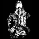
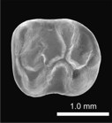
A new and primitive species of Protophiomys (Rodentia, Hystricognathi) from the late middle Eocene of Djebel el Kébar, Central TunisiaLaurent Marivaux
Published online: 02/06/2014 |
|

|
Un giraffidae dans le pliocène de Montpellier ?Claude GuérinPublished online: 31/10/1986Keywords: Artiodactyla; France; Giraffidae; Mammalia; Montpellier; Ruscinian https://doi.org/10.18563/pv.16.3.185-189 Abstract An upper giraffid premolar without any indication about its origin is preserved at the Montpellier University among numerous fossils from the ruscinian formation of Montpellier. It can be related to Samotherium, of the Upper Miocene in Eastern Europe, North Africa and Asia, or more probably to Bramatherium or Hydaspitherium of the Pliocene of South East Asia. The sedimentological study of the matrix shows a calcareous background, which may indicate that this tooth does not come from the Montpellier formation. PV article infos Published in Vol. 16, Fasc. 3 (1986) |
|

|
Le genre Microstonyx en Espagne et ses relations avec les autres espèces du même genre hors d'EspagneJuana M. Golpe-PossePublished online: 01/10/1980Keywords: Microstonyx; Spain; Suidae https://doi.org/10.18563/pv.9.ext.213-231 Abstract The genus Microstonyx was found only in the north eastern part of Spain : M. antiquus, referable to the PV article infos Published in Vol. 9, Ext (1980) |
|

|
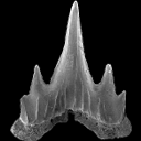
Hexanchiforme nouveau (Neoselachii) du Crétacé inférieur du Sud de la FranceHenri Cappetta
Published online: 18/12/1990 |
|