
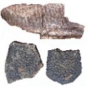
Pterosaurs from Coahuila
Abstract book of the 18th Conference of the EAVP
Two enigmatic rodents from Lavergne (MP 16), Quercy Phosphorites
Les sélaciens du Miocène de la région de Montpellier
Muridae du Pliocène supérieur d'Espagne et du midi de la France.
Les Chiroptères du Miocène inférieur de Bouzigues. 1- Etude systématique.
Eocene (57) , Quercy Phosphorites (38) , Systematics (32) , Rodents (29) , Mammalia (27) , Rodentia (25) , Miocene (24)

|
Field trip guides of the 20th Annual Conference of the European Association of Vertebrate Palaeontologists, 26th June – 1st July 2023, Sabadell (Barcelona), Spain
|
|

|
A new Ardynomys (Rodentia,Cylindrodontidae) from the Eocene of the eastern Gobi Desert, Mongolia.Demberelyin DashzevegPublished online: 16/12/1996Keywords: Ardynomys; Eocene; Mongolia; Rodentia; Systematics Abstract A partial skull of Ardynomys russelli sp. nov. (Rodentia, Cylindrodontidae) is described. This was collected in the late Eocene of Alag Tsab locality in the eastem Gobi Desert, Mongolia. Ardynomys russelli sp. nov. is characterized by small size, brachyodont molars, and retention of P3. It represents the earliest record of the genus Ardynomys MATTHEW & GRANGER, 1925, in Asia. PV article infos Published in Vol. 25, Fasc. 2-4 (1996) |
|

|
Diversity among north african dinosaur eggshells.Monique Vianey-Liaud
Published online: 15/12/2003 |
|
|
Terrestrial vertebrate paleocommunities from the Cerro del Pueblo Formation (Late Cretaceous; Late Campanian) at Las Aguilas, Coahuila, MexicoHéctor E. Rivera-Sylva
Published online: 16/07/2019 |
|

|
Contributions à l'étude du gisement Miocène supérieur de Montredon (Hérault). Les grands mammifères. 8 - Analyse paléoécologique de la faune mammalienneSerge Legendre
Published online: 15/11/1988 |
|

|
Rongeurs (Mammalia, Rodentia) du Miocène de Beni-MellalJean-Jacques Jaeger
Published online: 15/02/1977 |
|

|
Le genre Leptolophus (Perissodactyla, Mammalia): morphologie et histologie dentaires, anatomie cranienne, implications fonctionnelles.Jean-Albert RemyPublished online: 15/09/1998Keywords: dental histology; Eocene; functional anatomy; Palaeotheriidae; skull anatomy; Southern France; Systematics Abstract A strong lophodonty, an extreme heterodonty, some hypsodonty and regular overlayings of coronal cement are prominent features of the genus Leptolophus (Palaeotheriinae = Palaeotheriidae s.s.). The histological pattern of the teeth unusually joins type II enamel prisms, characteristic of advanced ungulates, together with archaic features, such as an almost complete lack of Hunter-Schreger zonation and a weak expanse of peritubular dentine. The skull is narrow and slender, with an elongated ante-orbital facial region, a moderately notched nasal aperture, a rather elongated post-canine diastem, parallel zygornatic arches and a fairly dorsally located squamoso-mandibular joint.The functional analysis brings to light "ectolophodont" masticatory cycles with two phases, in which maximum power was applied, contrary to equíds, on hindmost teeth; likewise, skull accomodations to increasing height of the teeth are quite different. This study leads to the assumption that Leptolophus may have been light mammals, living in rather open surroundings, browsing on herbaceous plants or leaves cropped close to the ground. Moreover, it appears that it could have been some inadequacy of dental structures to the dietary, which leaded to quick wear of the teeth and to many enamel notches, but had been somewhat balanced by the early increase of hypsodonty, not induced in such a case by a biotop deterioration (as it will happen at the end of the Eocene). This ínadaptation might account for the short duration of the genus Leptolophus, whose the 3 species, L. stehlini, L. nouletí and L. magnus n. sp. are indeed confined in the level MP 16. Its geographical spreading (as far as known, South of western Europe) and the morphological pattern of its dentition suggest that this genus would have been related to early upper Eocene endemic spanish forms. PV article infos Published in Vol. 27, Fasc. 1-2 (1998) |
|

|
Insectivores pliocènes du Sud de la France (Languedoc-Roussillon) et du Nord-Est de l'Espagne.Jean-Yves CrochetPublished online: 31/10/1986Keywords: Biostratigraphy; Insectivora; Languedoc; Pliocene; Spain; Systematics https://doi.org/10.18563/pv.16.3.145-171 Abstract The first lists of Insectivores (Erinaceidae, Talpidae and Soricidae) from the Pliocene beds of Southern France and North-East Spain are given in this paper. The material from twelve localities is studied. These localities are geographically situated in Languedoc (Celleneuve, Vendargues, Nîmes, Sète, Balaruc 2 and Seynes), in Roussillon (Terrats, Serrats-d'en-Vacquer, Château d'eau and Mont-Hélène) and in North-East Spain (Layna, Medas Islands and Puebla de Valverde). These faunas correspond to the Early, Middle and Late Pliocene. 1 to 8 taxa are identified in these localities and 14 specific taxa are presently listed for this period in this area. Two new specific taxa are described as Galerix depereti nov. sp. from all the Early Pliocene localities in the North-Pyrenean area and as Desmanella gardiolensis nov. sp. from Balaruc 2. For this small mammals, two faunal assemblages are recognized. The first one is dated from the Early Pliocene (F 1, 2 and 3 zones in Aguilar et Michaux) and is characterized by Galerix depereti and rare and little diversified Soricids. The second one is Late Pliocene in age (zones G 2 and G 3). The fossils of the genus Talpa are relatively abundant and the Soricids are diversified and very abundant. The Middle Pliocene (zone G 1) is a transitional period. ln these faunas, most of the insectivore genera are known from the European Late Miocene beds (8 on 10). This fact demonstrates a relative continuity between the invectivore faunas from the Late Miocene to the Early Pliocene. In conclusion, somme paleoecological considerations are suggested. PV article infos Published in Vol. 16, Fasc. 3 (1986) |
|

|
Les Gliridés (Rodentia) de l'Oligocène supérieur de Saint-Victor-la-Coste (Gard).Marguerite HugueneyPublished online: 28/10/1968Keywords: Gliridae; Late Oligocene https://doi.org/10.18563/pv.2.1.1-16 Abstract The locality of St.-Victor-la-Coste (Gard) has yielded, rather abundantly, teeth of two glirids hitherto very poorly known: Glirudinus praemurinus (Freudenberg) and Glirudinus glirulus (DEHM). It has permitted, moreover, new views on the evolution of Peridyromys murinus (POMEL). Study of these forms confirms the late Oligocene age of the fauna, without allowing, however, further precision. PV article infos Published in Vol. 02, Fasc. 1 (1968) |
|

|
First evidence of an early Miocene marine teleostean fish fauna (otoliths) from la Paillade.(Montpellier,France)Bettina Reichenbacher
Published online: 15/06/1999 |
|

|
Les mammifères Montiens de Hainin (Paléocène moyen de Belgique) Part III : MarsupiauxJean-Yves Crochet and Bernard SigéPublished online: 30/09/1983Keywords: Belgium; Marsupials; Paleobiogeography; Paleocene https://doi.org/10.18563/pv.13.3.51-64 Abstract The oldest european marsupials are described from some specimens (isolated upper molars) recently found from the Hainin sediment (Middle Paleocene of Belgium). These fossils document a new species of the Peradectes genus. They give evidence of a much older occurrence of the marsupials in Europe than it was assumed. They allow us to postulate a didelphid dispersal from South America towards the western-holarctic area operating in two phases : the first one of the Peradectes genus at the end of the Cretaceous; the second one of the Didelphíni tribe at the end of the Paleocene. A central american crossing is likely for the first one, whereas a transafrican way is tentatively argued for the second one. PV article infos Published in Vol. 13, Fasc. 3 (1983) |
|

|
Les rongeurs du Miocène moyen et supérieur du MaghrebJean-Jacques Jaeger
Published online: 15/05/1977 |
|
|
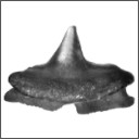
Batoids (Rajiformes, Torpediniformes, Myliobatiformes) from the Sülstorf Beds (Chattian, Late Oligocene) of Mecklenburg, northeastern Germany: a revision and description of three new speciesThomas ReineckePublished online: 24/06/2015Keywords: Batoids; Chattian; Elasmobranchii; North Sea Basin; Oligocene https://doi.org/10.18563/pv.39.2.e2 Abstract Bulk-sampling of fossil-rich tempestites from the Chattian Sülstorf Beds of PV article infos Published in Vol.39-2 (2015) |
|

|
Perutherium altiplanense, un Notongulé du Cretacé Supérieur du PérouLarry G. Marshall, Christian de Muizon
Published online: 30/11/1983 |
|

|
Observations sur l'anatomie crânienne du genre Palaeotherium (Perissodactyla, Mammalia): mise en évidence d'un nouveau sous-genre, FranzenitheriumJean-Albert RemyPublished online: 01/12/1992Keywords: Palaeotherium; Paléogène; Perissodactyla; skull anatomy; Systematics Abstract The skull remains referred to the genus Palaeotherium are recorded and described. Biometrical tests are made to elucidate intrageneric allometric relationships and to allow comparisons with various other perissodactyls. Apart from the well known shortness of post canine diastems and deepness of the narial opening, the genus is characterized by a great lengthening of the splanchnocranium, owing to a spreading of the post-orbital facial region, by a reduced area of the eye-socket and by the prevalence of the temporal muscle with regard to the masseter; this original shape of the masticatory apparatus needs to be related to the morphology of the jugal teeth and particularly to their asymmetrical semi-hypsodonty. PV article infos Published in Vol. 21, Fasc. 3-4 (1992) |
|

|
The skull of Arsinoitherium (Mammalia, Embrithopoda) and the higher order interrelationships of ungulatesNicholas CourtPublished online: 17/12/1992Keywords: Arsinoitherium; PHYLOGENY; Skull; Ungulate Abstract Detailed anatomical description of arsinoithere cranial remains from the Lower Oligocene, Fayum Depression, Egypt, provides the basic data for a systematic investigation. All cranial and some postcranial features are assessed from a phylogenetic standpoint. Several soft tissue characters are then added to a cladistic analysis based on 54 derived ungulate morphological characters. The resulting phylogenetic hypothesis implies that perissodactyls, sirenians, proboscideans and arsinoitheres constitute a monophyletic unit (5 synapomorphies). However, increasing the tree length by 3 steps reveals a closer association between hyraxes and perissodactyls. Nevertheless, 13 synapomorphies link proboscideans, sirenians and arsinoitheres to the exclusion of all other ungulates. Form of the sphenopalatine and ethmoid foramina, recurved posttympanic process, absence of a fenestra rotundum in the petrosal, vestigial paroccipital process of the exoccipital and the highly unusual absence of a hypoglossal foramen in the skull, imply a robust sister-group relationship between arsinoitheres and proboscideans. In this analysis artiodactyls share only one derived character with all other ungulates studied. Monophyly of Ungulata, including Artiodactyla, is therefore only weakly supported. It is argued that pedal anatomy of hyraxes is non-homologous with that of Tethytheria. Arsinoitherium should now be classified within Tethytheria, sharing a sister-group relationship with Proboscidea. Hyraxes are excluded, thus refuting the concept of Paenungulata. However, monophyly of the wider concept, Pantomesaxonia, containing hyraxes, perissodactyls, sirenians, proboscideans and now, arsinoitheres, is supported by this study. PV article infos Published in Vol. 22, Fasc. 1 (1992) |
|
|
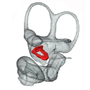
Fallen in a dead ear: intralabyrinthine preservation of stapes in fossil artiodactylsMaeva J. Orliac
Published online: 09/03/2016 |
S.I. Data |

|
Henri Menu, 1925-2007Bernard SigéPublished online: 16/12/2008Keywords: bats; biography https://doi.org/10.18563/pv.36.1-4.1-5 Abstract Record of life and works of Henri Menu, French zoologist, contributor to the knowledge of living and fossil bats. PV article infos Published in Vol. 36, Fasc. 1-4 (2008) |
|

|
Révision des Rhombodontidae (Neoselachii Batomorphii) des bassins à phosphate du MarocAbdelmajid Noubhani and Henri Cappetta
Published online: 20/05/1994 |
|
|
New Late Miocene plecotine bats (Chiroptera, Vespertilionidae: Plecotini) from Gritsev, UkraineValentina V. Rosina, Sergei Kruskop
Published online: 07/03/2019 |
|