
|
Mammifères de l'Ilerdien Moyen (Eocène inférieur) des Corbières et du Minervois (Bas-Languedoc, France). Systématique, Biostratigraphie, Corrélations.
Published online: 25/01/1991
Keywords:
Biostratigraphy; Corbières; correlations; Early Eocene; Ilerdian; Mammalia; Minervois; Paleobiogeography; Southern France
Abstract
Mammal-bearing localities have been discovered in the marine and lacustrine series of the middle Ilerdian (Lowermost Eocene) from Southem France (Minervois and Corbières). In the localities of Fordones, Monze, Fournès, and La Gasque, thirty mammal species have been identified. Among others, they include ischyromyid rodents (Microparamys and Pseudoparamys), paromomyid and adapid primates (Arcius and Donrussellia), new insectivores, condylarths, and a dyspternine pantolestid. These faunas provide new informations on the early Eocene Mesogean faunas of Rians and Palette. The assemblages of primates and rodents from Fordones support good correlations with Palette which was recently placed near the standard-level of Dormaal (MP 7). In fact, Palette and Fordones could be even older than Dormaal. Consequently, there seems to be a relatively important temporal gap between the late Paleocene of Cernay and the Sparnacian of Dormaal. This gap could be partly filled with the Mesogean faunas of Palette, Fordones, and Silveirinha. On the basis of these new mammal faunas the marine middle Ilerdian is proved to be older than the Cuisian stage of the Paris Basin. With regards to the position of the Fordones fauna at the top of the NP 10 calcareous nannoplankton biozone, the westem European paleomammalogists Paleocene/Eocene boundary could be situated between the NP 9 and NP 10 biozones.
PV article infos
Published in Vol. 20, Fasc. 2-3 (1991)
|
PDF
|
|
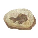
Designating a lectotype for Mesacanthus pusillus (Gnathostomata: Acanthodii)
Published online: 03/03/2021
Keywords:
acanthodians; Chordata; Devonian; Midland Valley; Orcadian Basin
https://doi.org/10.18563/pv.44.1.e2
Abstract
The early gnathostome genus Mesacanthus is well represented in both Lower Old Red Sandstone and Middle Old Red Sandstone assemblages of northern and central Scotland. This ‘acanthodian’ taxon is currently thought to comprise two valid species: M. mitchelli and M. pusillus. Although the whereabouts of the holotype of M. mitchelli (NHMUK PV P560) is known, the syntype material for M. pusillus has long been thought lost. Here we identify at least one specimen that formed part of the original syntype material for M. pusillus, albeit in a slightly different condition than when it was originally figured. This specimen is ROM 25872, which is here designated as the lectotype. A second specimen – ELGNM 1978.191.1 – could represent another of the syntype specimens, but poor preservation quality makes it impossible to be certain.
PV article infos
Published in 44-1 (2021)
|
PDF
S.I. Data
|

|
Evolution des Aplodontidae Oligocènes Européens
Published online: 01/10/1979
Keywords:
Aplodontidae; Europe; Oligocene
https://doi.org/10.18563/pv.9.2.33-82
Abstract
Until now Aplodontidae of the European Oligocene have been documented by four species only. The phylogenetic relations remained obscure. as the distribution of only one species has been known in some detail. New material made it possible to define the stratigraphic range of two of the already existing species (Plesispermophilus angustidens, Sciurodon cadurcense) and to follow their development during the Oligocene beginning with the event of the « Grande Coupure ››. Sciurodon remained nearly without change until the end of the Middle Oligocene. Plesispermophilus angustidens split into two distinct phyletic lines, one of which (P. macrodon n. sp.) reaching considerable size, is represented till the beginning of the Upper Oligocene (Pech de Fraysse, Gaimersheim). The other line leads to Plesispermophilus ernii (basal Upper Oligocene of Burgmagerbein 1. terminal Upper Oligocene of Coderet). Besides the already known forms a new small-sized species (P. atavus n. sp.) is described, which by its primitive features closely resembles the genus Plesispermophilus. Two other small-sized species already known from the Upper Oligocene (? P. argoviensis) and Lower Miocene (? P. descedens) seem to be closely related to the new species. It cannot be decided whether they are descendents of this line or have developed independently, because of their poor fossil record.
Comparison of the evolutionary modalities in the different phylogenetic lines reveals general trends. the most striking of which is the complication of the tooth pattern by the development of additional crests. In the lower molars the cusps diminuate in size and are more and more transformed into ridges. ln addition new connection between the crests appear. in the upper molars, the « selenodont » shape of the teeth becomes more and more dominant, and in the two main evolutionary lines of Plesispermophilus the metaconulus becomes duplicated. A further evolutionary trend is the size increase of the premolars compared to the molars, which is even more pronounced in the Miocene Aplodontidae.
Phylogenetic relations between the primitive Plesispermophilus and certain « prociurines ›› of Northern America as well as between Plesispermophilus (P. angustidens) and more progressive forms of the Upper Oligocene (P. ernii, P. macrodon n. sp.) can be documented. In this light, the taxonomic distinction between Prosciurinae (bunodont) and Allomyinae (selenodont) sensu Rensberger 1976 can be shown to be artificial, because it separates forms from each other, which are evidently closely related. Consequently the separation into two subfamilies has been abolished.
PV article infos
Published in Vol. 09, Fasc. 2 (1979)
|
PDF
|

|
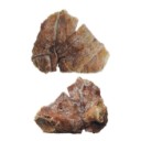
Nouveau Dichobunidae (Artiodactyla, Mammalia) du gisement d'Aumelas (Hérault) d'âge Lutétien terminal
Published online: 01/10/1980
Keywords:
Aumelas; Dichobunidae; Hérault; Middle Eocene; Upper Lutetian
https://doi.org/10.18563/pv.9.ext.197-211
Abstract
The faunal list of the mammals collected at the locality of Aumelas (Hérault, France) is revised. For the first
time this Middle Eocene locality is precisely settled in the european chronological scale of "niveaux repères", between the levels of Bouxwiller and Egerkingen, in Uppermost Lutetian.
A new Dichobunid from the site is described : Aumelasia gabineaudi n. g., n. sp. This new genus has primitive characters. and it may be in the descent of the Lower Eocene Protodichobune.
PV article infos
Published in Vol. 9, Ext (1980)
|
PDF
|

|
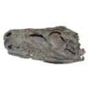
Crivadiatherium iliescui n. sp., nouvel Embrithopode (Mammalia) dans le Paléogène ancien de la dépression de Hateg (Roumanie).
Published online: 30/12/1985
Keywords:
Embrithopods; Late Eocene; Paleobiogeography; Romania
https://doi.org/10.18563/pv.15.3.139-157
Abstract
The investigations undertaken at Crivadia (Hateg Depression, Hunedoara District, Romania), the type locality of Crivadiatherium mackennai RADULESCO el al. (Radulesco, Iliesco et lliesco, 1976), led to the discovery of remains of a new Embrithopod. Close to the above mentioned species, but larger in size, this animal is here described as a new species of Crivadiatherium, C. iliescui. ln addition, the comparison made between the forms indicated above and Palaeaamasía kansui OZANSOY from the Eocene deposits of Anatolia (Ozansoy, 1966; Sen et Heintz, 1979) showed that the latter species included a heterogeneous material; this permitted us to distinguish the form in the Anatolian locality Ciçekdag-Arabin Kôyü under the name Palaeoamasia sp. The geographical distribution and diversity of the Embrithopod species under discussion (Balkan, Anatolia) support the idea of an eurasiatic origin of this group and seem to suggest the existence during the Eocene of a particular faunal province in south-eastern Europe.
PV article infos
Published in Vol. 15, Fasc. 3 (1985)
|
PDF
|

|
Les mammifères Montiens de Hainin (Paléocène moyen de Belgique) Part II : Les Condylarthres
Published online: 30/12/1982
Keywords:
Belgium; Condylarths; Louisininae; Oxyclaeninae; Paleocene
https://doi.org/10.18563/pv.12.6.173-184
Abstract
The Condylarths from Hainin (Hainault, Belgium) show no affinity at the generic level to those known in other Paleocene localities of Europe and North America ; they are described as new forms : Monshyus praevius n. gen., n. sp. and Prolatidens waudruae n. gen., n. sp. Monshyus praevius, discovered in only one of the levels in the excavation at Hainin, is similar to the genera Microhyus TEILHARD and Louisina RUSSELL ; with them it is included in the subfamily Louisininae (Hyopsodontidae). With respect to Microhyus and Louisina, Monshyus is distinguished by the precociously modern aspect of its upper molars, the only teeth that are referable. Prolatidens waudruae, known only by lower molars, was found in several levels in the pit at Hainin. It is an arctocyonid presenting possible relationships to the North American form Oxyprimus galadrielae ; it therefore has been provisionally attributed to the subfamily Oxyclaeninae. If this attribution is confirmed, this species will constitute the first and only representative of the group in Europe.
PV article infos
Published in Vol. 12, Fasc. 6 (1982)
|
PDF
|
|
Difficulties with the origin of dinosaurs: a comment on the current debate
Published online: 01/07/2020
Keywords:
dinosaur anatomy; dinosaur evolution; Ornithoscelida; palaeobiogeography; Triassic Period
https://doi.org/10.18563/pv.43.1.e3
Abstract
The origin and early evolutionary history of the dinosaurs is a topic that has recently gone through a period of renewed interest and academic debate. For 130 years, one way of classifying the various dinosaur subgroups persisted as the accepted model, with increasing levels of research in the past quarter-century also providing evidence for the hypothesis that dinosaur origination occurred in the Southern Hemisphere, particularly in South America. It is, after all, from within the Late Triassic strata of countries like Argentina and Brazil that we get some of the very best early dinosaur specimens; many of these specimens are the earliest known representatives of some of the major dinosaur subgroups, such as the theropods and sauropodomorphs. However, some recent analyses have brought about a shift in terms of what is currently accepted and what is now disputed regarding the origin of dinosaurs – the Southern Hemisphere origination hypothesis was questioned (although this was based upon observations and not with quantitative analysis techniques), as has the shape of the dinosaur tree. Responses to the new hypothesis were numerous; many further supported a Southern Hemisphere point of origin. Whilst the interrelationships between the major dinosaur clades remains to be resolved, the current data does seem to comprehensively answer the question of where the dinosaurs first originated. However, it is arguable whether the current data that is being used in such palaeobiogeographical analyses is sufficient to provide an answer to the question of where specifically the dinosaur clade first appeared. This short communication urges a degree of caution about the current consensus and what steps may need to be taken to ensure that more meaningful results are produced in the future.
PV article infos
Published in Vol 43-1 (2020)
|
PDF
|

|
Leptacodon nascimentoi n,sp., un nouveau Nyctitheriidae (Mammalia,Lipotyphla) de l'Eocène inférieur de Silveirinha (Baixo Mondego, Portugal)
Published online: 16/12/1996
Keywords:
Eocene; Leptacodon; Lipotyphla; Mammals; Nyctitheriidae; Portugal; Silveirinha
Abstract
In this article is described a new species of Nyctitheriidae with primitive characters: Leptacodon nascimentoi n. sp. from the early Eocene of Silveirinha (Portugal).
PV article infos
Published in Vol. 25, Fasc. 2-4 (1996)
|
PDF
|

|
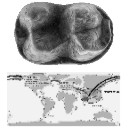
An Australian Miocene Brachipposideros (Mammalia, Chiroptera) related to Miocene representatives from France
Published online: 15/12/1982
Keywords:
Australia; bats; Chiroptera; Miocene
https://doi.org/10.18563/pv.12.5.149-172
Abstract
A new middle Miocene hipposiderid bat is described from a limestone deposit on Riversleigh Station in north-western Queensland. Hipposideros (Brachipposideros) nooraleebus n. sp. is the first record of this subgenus from anywhere in the world outside of France. The palaeoecological setting of the fossil bats appears to have been a relatively quiet, sunny lime-enriched tropical pool that contained tortoises, crocodiles and fish. It is possible that the bats were washed into the pool from an adjacent cave.
The Riversleigh bat most closely resembles the French Burdigalian (early middle Miocene) bat H. (B.) aguilari. It is also possible that it may have been closely related to the original Australian hipposiderid stock that ultimately gave rise to the endemic monotÿpic Rhinonycteris aurantius. The disjunct distribution of species of H. (Brachipposideros) suggests that representatives of this subgenus will be found in at least tropical southern Asia.
PV article infos
Published in Vol. 12, Fasc. 5 (1982)
|
PDF
|

|
Analyse d'ouvrage : A.J.Sutcliffe. On the track of Ice Age mammals
Published online: 30/12/1985
Keywords:
Book review
https://doi.org/10.18563/pv.15.4.270
Abstract
On the Track of Ice Age Mammals justifie pleinement son titre car l'auteur, Anthony J. Sutcliffe, apporte au lecteur faits et interprétations qui l'amèneront à s'intéresser encore plus au passé récent et à l'avenir de son environnement et à la question de l'impact de l'homme sur la nature. Après les chapitres qui présentent les temps glaciaires et les divers témoignages qui nous en sont parvenus, les cinquième et sixième apportent les informations nécessaires à la compréhension des résultats que nous donnent les chercheurs: principes, moyens d'étude et limites des méthodes, difficultés de l'intégration des données dans un cadre stratigraphique, variabilité des signaux climatiques, variabilité de leur intensité selon l'endroit par rapport au centre de la glaciation, complications liées à la qualité inégale de l'enregistrement géologique, en mer et sur le continent.
PV article infos
Published in Vol. 15, Fasc. 4 (1985)
|
PDF
|

|
The eosimiid and amphipithecid primates (Anthropoidea) from the Oligocene of the Bugti hills (Balochistan, Pakistan): new insight into early higher primate evolution in South Asia.
Published online: 15/10/2006
Keywords:
Amphipithecidae; anthropoid phylogney; Bugti Hills; Early Oligocene; Eosimiidae; Pakistan
https://doi.org/10.18563/pv.34.e15
Abstract
Eosimiid and amphipithecid primates document a long and significant history of primate evolution throughout the Eocene in Southeast Asia. Despite the absence of a comprehensive post-Eocene fossil record, it was generally hypothesized that both families left no descendant in Asia. Recently, two new small-bodied taxa, Bugtipithecus and Phileosimias, have been recovered in early Oligocene coastal deposits from the Bugti Hills (Balochistan, central Pakistan) and referred to the families Amphipithecidae and Eosimiidae, respectively, on the basis of dental fossil remains. In this paper, we provide more exhaustive description, comparison, and discussion of these taxa. As for tarsiid and sivaladapid primates, the persistence of eosimiids and amphipithecids into the Oligocene clearly demonstrates that low latitudes of South Asia provided a continuous access to tropical refugia during the climatic deterioration characterizing the late Eocene-early Oligocene interval, which was seemingly lethal for primate communities elsewhere across the Holarctic continents. As a contribution to the ongoing phylogenetic debates regarding the position of eosimiids and amphipithecids on the primate family tree, we have performed a cladistic analysis in a high-level primate systematic context in order to assess the position and the role of these new taxa in that phylogenetic issue. Our results support the view according to which eosimiids and amphipithecids (and by extension Phileosimias and Bugtipithecus, respectively) are stem anthropoids. These fossils from Pakistan document an unsuspected Oligocene phase of the evolutionary history of anthropoid primates in southern Asia, which clearly enhances the extent of the anthropoid radiation in this province during the Paleogene. Several phylogenetic and paleobiogeographic aspects are discussed, notably the intra- and inter-relationships between Paleogene Asian and Afro-Arabian anthropoids, and the resulting potential dispersal models between both land-masses during the Paleogene.
PV article infos
Published in Vol. 34, Fasc. 1-2 (2006)
|
PDF
|

|
Les rongeurs de l' Eocène inférieur et moyen d'Europe Occidentale; Systématique, phylogénie, biochronologie et paléobiogéographie des niveaux-repères MP 7 à MP 14.
Published online: 15/12/1999
Keywords:
Biochronology; Early and Middle Eocene; Gliridae; Ischyromyidae; Mammalia; MP Scale; New Genus and Species; Palaeogeography; PHYLOGENY; Rodents; Theridomyidae; Western Europe
Abstract
Fourteen distinct phyletical lineages which belong at least in three families: Ischyromyidae ALSTON, 1876, Gliridae THOMAS, 1896 and Theridomyidae ALSTON, 1876, have been identified after the study of more than 3600 rodent dental remains from about twenty Early and Middle Eocene european localities. A systematical and phylogenetical revision of these rodents has been achieved. Nearly all the specific and generic diagnosis are emended. Several new combinations and synonymies are proposed. Four new species and two new genera, Euromys nov. (Ailuravinae) and Hartenbergeromys nov. (Microparamyini), are named and described. Euromys nov. gen. is known by three distinctive ypresian (MP 7 to MP 10 european reference levels) chronospecies. This new lineage is thought to be the direct ancestor of Meldimys MICHAUX, 1968 and Ailuravus RUTIMEYER, 1891. A new species of the genus Plesiarctomys BRAVARD, 1850, Pl. lapicidinarum from Condé-en-Brie (MP 8-9 reference level), allows to relate the Plesiarctomys lineage to the Pseudoparamys MICHAUX, 1964 one. The taxa Sparnacomys HARTENBERGER, 1971, Pantrogna HARTENBERGER, 1971, and Corbarimys MARANDAT, 1989 are erected to genus rank; the last one is not thought to be an Ischyromyidae. A new chronospecies of Pantrogna, P. marandati nov. sp. from the locality of Prémontré (MP 10 reference level), is described. This lineage is at the origin of two others, namely Masillamys TOBIEN, 1954, including M. mattaueri (HARTENBERGER, 1975) nov. comb. (MP 10 reference level), and Hartenbergeromys nov. gen., known from MP 10 (H. hautefeuillei nov. sp.) and MP 11 (H. parvus TOBIEN, 1954) reference levels. The phylogenetical position of Hartenbergeromys nov. gen., at the origin of the european family Theridomyidae, is discussed. The systematical and phylogenetical status of two probable Paramyinae, "Paramys" woodi MICHAUX, 1964 and an unnamed genus and species, are discussed. New populations of the primitive Gliridae Eogliravus HARTENBERGER, 1971 and of the primitive Theridomyidae Protadelomys HARTENBERGER, 1968, are described and assigned to previously known species.
PV article infos
Published in Vol. 28, Fasc. 2-4 (1999)
|
PDF
|
|
First early Eocene tapiroid from India and its implication for the paleobiogeographic origin of perissodactyls
Published online: 08/09/2015
Keywords:
Ceratomorpha; Helaletidae; Paléogène; Tapiromorpha; Vastan
https://doi.org/10.18563/pv.39.2.e5
Abstract
The presence of cambaytheres, the sister group of perissodactyls, in western India near or before the time of collision with Asia suggests that Perissodactyla may have originated on the Indian Plate during its final drift towards Asia. Herein we reinforce this hypothesis by reporting two teeth of the first early Eocene tapiromorph Perissodactyla from the Cambay Shale Formation of Vastan Lignite Mine (c. 54.5 Ma), Gujarat, western India, which we allocate to a new genus and species, Vastanolophus holbrooki. It presents plesiomorphic characters typical of the paraphyletic “Isectolophidae,” such as small size and weak lophodonty. However, the weaker hypoconulid and low paralophid, higher cusps, lower cristid obliqua, and the lingual opening of the talonid are found in Helaletidae, the most primitive tapiroid family. V. holbrooki, gen. et sp. nov., may be the oldest and the most primitive tapiroid, suggesting that at least tapiroid perissodactyls originated on India.
PV article infos
Published in Vol.39-2 (2015)
|
PDF
|

|
Koobi Fora Research Project, volume 3. The fossil ungulates: geology, fossil artiodactyls, and palaeoenvironments, édité par John Michael HARRIS, 1991. Clarendon Press, Oxford, xvi + 384 p. ISBN 0-19-857399-5.
Published online: 11/02/1993
Keywords:
Artiodactyls; palaeovenvironments; Ungulates
Abstract
Avec ce volume se clôture l'étude géologique et paléontologique des sites à hominidés de Koobi Fora. Il fait suite aux deux précédents ouvrages, parus respectivement en 1978 et 1983, consacrés également à l'étude des faunes recueillies sur ces gisements dans le cadre du Koobi Fora Research Project.
PV article infos
Published in Vol. 22, Fasc. 2-3 (1993)
|
PDF
|
|
Dortokid turtle remains from the Upper Cretaceous of Cruzy (Hérault, southern France) and phylogenetic implications
Published online: 14/11/2022
Keywords:
Cruzy; Dortoka vasconica; France; Late Cretaceous; Turtle
https://doi.org/10.18563/pv.45.2.e3
Abstract
An isolated right costal 1 from the Late Cretaceous Massecaps locality (Cruzy, Hérault, southern France) is assigned to Dortoka vasconica (Dortokidae). This find adds a new element to the Late Cretaceous turtle fauna of Cruzy and further supports the hypothesis that two distinct lineages of Dortokidae were present in Europe during the Late Cretaceous-Paleogene due to geographical isolation.
PV article infos
Published in 45-2 (2022)
|
PDF
|

|
This review critiques the third edition (1986) of The Animals (Systematische Zoologie), a descriptive zoology textbook first published in 1976. The book focuses on morphology, taxonomy, and basic anatomy of animal groups, with occasional notes on distribution, habits, and economic/medical significance.
Published online: 15/04/1986
Keywords:
Book review; Systematics
https://doi.org/10.18563/pv.16.1.55
Abstract
THE ANIMALS. Systematische Zoologie by A. REMANE, V. STORCH, and U. WELSCH. 1986, Edition 3. Cloth and paperbound. Stuttgart and New York : Gustav Fischer Verlag. xvi + 698 pp. DM 86. (Geb.) & DM 72 (kart.).
PV article infos
Published in Vol. 16, Fasc. 1 (1986)
|
PDF
|

|
Lower Paleogene crocodilians from Silveirinha, Portugal.
Published online: 15/10/2003
Keywords:
?Upper Paleocene / Lowermost Eocene; Crocodilians; Ecology; Portugal
Abstract
The presence at Silveirinha of one of the earliest, ? Late Paleocene or Lowermost Eocene, european representatives of the genus Diplocynodon is based mostly on isolated bones and teeth (often from juveniles). This small-sized form is the only crocodilian so far recognized in this site. The longevity of Diplocynodon in Portugal becomes much extended; the genus survived there until the Middle Miocene at least. A discussion on the possible affinities with other eocene Díplocynodon and especially those from Cubillos-Valdegallina (Zamora, Spain) is presented. On the other hand, differences have been detected in comparison with: Díplocynodon tormis, from the middle Eocene of the Douro basin in Spain, which may belong to another phyletic line; and the aff. Diplocynodon from Dormaal (Belgium) and Le Quesnoy (France), nearly contemporaneous of Silveirinha. The Silveirinha Diplocynodon and many other data strongly suggest moist, subtropical, quite limited in space environments related to an alluvial plain crossed by small, meandering channels.
PV article infos
Published in Vol. 32, Fasc. 1 (2003)
|
PDF
|

|
Etude des dents jugales inférieures des Equus (Mammalia, Perissodactyla) actuels et fossiles
Published online: 15/04/1981
Keywords:
Cheek teeth; Equus; Mammals
https://doi.org/10.18563/pv.10.3-4.127-226
Abstract
The comparative morphology and biometry of the lower cheek teeth of modern Equus are studied on approximately 300 mandibles belonging to the 10 usually recognised species : Equus grevyi, E. burchelli, E. quagga, E. zebra, E. africanus, E. asinus, E. hemionus, E. klang, E. przewalskii, E. caballus. The studied parameters comprise : occlusal length and width, postflexid length and index ; shape of the double knot (metaconid + metastylid + lingual groove) ; depth of the vestibular groove on the molars ; frequency of the pli caballinid, protostylid and other enamel plications or islets ; frequency of the dP/l.
The same methods of study are applied to a number of North American, Eurasian and African species. For the sake of comparison, some Hemphillian equids were observed (Dinohippus interpolatus, Dinohippus leidyanus, Astrohippus ansae, Phiohippus mexicanus) but most of the discussed material belongs to Pliocene or Pleistocene species of Equus : the « stenonine ›› E. stenonis, E. simplicidens, E. sanmeniensis and E. teilhardi; the « caballine ›› E. scotti, E. lambei, E. Iaurentius, E. mosbachensis, E. germanicus, E. gallicus, E. taubachensis and the Liakhov horse. The relationships of other species, in particular the North American E. calobatus, E. occidentalis, E. cf mexicanus are not clear for the moment. ln Africa, the Plio-Pleistocene species from Koobi Fora (Kenya) show some stenonine and perhaps asinine affinities. The relationships of E. numidicus and E. tabeti are uncertain but these species are probably related to the East African ones. E. mauritanicus is most certainly related to the Quagga group.
The biometrical data are gathered in 32 tables ; 4 photographie plates and 19 figures illustrate the next. The whole is a complement of the previously published studies of the skulls, upper cheek teeth, incisors and metapodials of modern and fossil Equus.
PV article infos
Published in Vol. 10, Fasc. 3-4 (1981)
|
PDF
|

|
Cervus elaphus rossii (Mammalia, Artiodactyla), a new endemic sub-species from the Middle Pleistocene of Corsica
Published online: 28/12/2001
Keywords:
Cervus elaphus; Corsica; Endemism; Pleistocene
Abstract
Several endemic deer remains from the Middle Pleistocene deposits of the Castiglione cave (Oletta, Haute-Corse) are examined here. A morphometric analysis allows to relate them to a new insular subspecies Cervus elaphus rossii. The bones were compared with those of the mainland early Middle Pleistocene subspecies Cervus elaphus acoronatus Beninde and the European species Cervus elaphus Linné (Late Middle Pleistocene and Upper Pleistocene forms (continental and insular)). The Castiglione fossil shows peculiar morphofunctional features in its appendicular skeleton suggesting a morphological convergence with certain Bovidae.
PV article infos
Published in Vol. 30, Fasc. 3-4 (2001)
|
PDF
|
|
First report of Cylindracanthus (Osteichthyes) from the Eocene of India
Published online: 25/03/2024
Keywords:
Cylindracanthus; Eocene; histology; rostrum; Umarsar mine.
https://doi.org/10.18563/pv.47.1.e2
Abstract
Fossils of the endangered sturgeons and peddlefishes are widely distributed. We here report for the first time the presence of one of the extinct osteichthyes genus Cylindracanthus (Liedy 1856a) from the Early Eocene lignite-bearing successions of the Kutch Basin, India. The present well preserved rostrum is characterised by numerous wedge-shaped components encircling the central canal that runs along its length, paired at the base and each wedge contributing to the formation of a ridge. The rostrum lacks teeth. The present find extends the palaeobiogeographical distribution of Cylindracanthus considerably and supports its Eocene age as dental remnants preserved in Cylindracanthus sp. shows a decrease in remanent dentition and tooth bases from the Cretaceous to the Eocene. Cylindracanthus is an useful palaeoenvironmental indicator as it has been found associated typically with deposits of nearshore marine environments.
PV article infos
Published in 47-1 (2024)
|
PDF
|
