
|
Contributions à l'étude du gisement Miocène supérieur de Montredon (Hérault). Les grands mammifères. 9 - Une tentative de magnétostratigraphie
Published online: 15/11/1988
Keywords:
France; magnetostratigraphy; Montredon; Upper Miocene
https://doi.org/10.18563/pv.18.ext.187-188
Abstract
Afin d'étudier la magnétostratigraphie de la colline de Montredon, j'ai prélevé en Avril 1982, 93 carottes orientées réparties sur 29 sites. J.-P. Aguilar et I. Loridant m'ont aidé durant cette opération. Pour la stratigraphie de la coupe échantillonnée et pour le contenu faunique des deux gisements de mammifères de cette colline, voir J.-P. Aguilar & J.-Y. Crochet (1982).
[...]
PV article infos
Published in Vol. 18, Ext (1988)
|
PDF
|

|
Strange Eocene rodents from Spain
Published online: 16/12/1996
Keywords:
Biogeography; Eocene; PHYLOGENY; Rodents; Spain; Zamoramys extraneus n. gen. n. sp.
Abstract
A new European rodent from the middle Eocene of Spain, Zamoramys extraneus n. gen., n. sp., appears to be closely related to the middle Eocene chapattimyid rodents of Indo-Pakistan. This contradicts the generally accepted paleobiogeographic hypothesis of a Tethyian barrier between Europe and Asia isolating Europe during the middle Eocene. Because of this barrier, some authors have proposed that European and Asian rodents were not closely related, their similarity being the result of morphological convergence. Here monophyly has been tested, using the parsimony criterion, based on an analysis of dental characters (including discussing of homology and the validity of some characteristics). Our results indicate a phylogenetic relationship among the Asiatic Ctenodactyloidea, Zamoramys from Spain, and the European endemic Theridomyoidea. We also conclude from our analysis that theridomyoids and European ischyromyoids are probably not closely related phylogenetically.
PV article infos
Published in Vol. 25, Fasc. 2-4 (1996)
|
PDF
|

|
New late Paleocene rodents (Mammalia) from Big Multi Quarry, Washakie Basin,Wyoming.
Published online: 16/12/1996
Keywords:
Clarkforkian; North America; Paleocene; Rodentia
Abstract
The earliest North American rodents occur in basal Clarkforkian beds of the Fort Union Formation at Big Multi Quarry near Bitter Creek, northern Washakie Basin, Sweetwater County, Wyoming, and in closely correlative Fort Union beds formerly accessible in the Eagle Coal Mine near Bear Creek, northern Clark's Fork Basin, Carbon County, Montana. Two new species of early Clarkforkian rodents, Paramys adamus and Alagomys russelli, are described from Big Multi Quarry. Paramys adamus is represented by virtually complete upper and lower dentitions, which demonstrate that this species is one of the most primitive North American paramyids yet discovered. These specimens form the basis for a reevaluation of the content and stratigraphic range of P. atavus, which is known with certainty only from Bear Creek. Alagomys russelli is the first North American record for the enigmatic rodent family Alagomyidae, otherwise known from ?late Paleocene-early Eocene localities in Mongolia and China. Phylogenetic analysis of dental and gnathic traits suggests that Alagomyidae form the sister group of all other undoubted rodents. At least two rodent clades, alagomyids and basal paramyids, seem to have invaded North America from Asia at the beginning of Clarkforkian time, but only the paramyids persisted to undergo a significant evolutionary radiation in North America.
PV article infos
Published in Vol. 25, Fasc. 2-4 (1996)
|
PDF
|

|
Un gisement à mammifères dans la formation lacustre d'âge Miocène moyen du Collet Redon près de St-Cannat (Bouches-du-Rhone). Implications stratigaphiques
Published online: 10/04/1979
Keywords:
France; Neogene; Rodentia
https://doi.org/10.18563/pv.8.5.327-341
Abstract
The new fauna of Collet Redon (Bouches-du-Rhône, France) is dated by three rodents: Megacricetodon aff. bavaricus, Democricetodon affinis mutilus and Peridyromys cf. hamadryas. They correlate this locality with Oggenhof and Ohningen in Bavaria (Western Germany). As the radiometric age of Ohningen is estimated between 14 and 13 M.Y., these three localities are of Serravallian age. This datation brings a complete readjusment of the stratigraphy of the section of Collet Redon formerly described by Collot and Combaluzier. The marine deposits with underly the continental formation with the mammal fauna, are Burdigalian. The angular unconformity between the marine and the continental deposits gives evidence of an episode of emersion on the margin of a sedimentary basin, with deformation and erosion. Owing to the newly discovered fauna, this geodynamical event is clearly settled within the regional geographical and chronological context. Lacustrine and continental deposits of such an age were up to now unsuspected in this area.
PV article infos
Published in Vol. 08, Fasc. 5 (1979)
|
PDF
|

|
Preliminary evolution of paleosols and implications for interpreting vertebrate fossil assemblages, Kuldana formation, Northern Pakistan
Published online: 16/12/1996
Keywords:
Eocene; Kuldana Formation; Pakistan; Paleosols; Pedogenic Carbonate; taphonomy; Time Averaging; Vertebrate Fossils
Abstract
Paleosols and the taphonomy of vertebrate fossils in the Eocene Kuldana Formation of northern Pakistan provide important information on the preservation and time-averaging of fossil assemblages. Morphologic, mineralogic, and chemical data as well as comparisons with Quaternary soils suggest that Kuldana paleosols formed under generally dry and oxidizing conditions over time intervals of less than 100 000 years and perhaps as short as 1000 years. The distribution of carbonate in Kuldana paleosols further indicates that the upper half of the profiles were acidic whereas the lower halves were alkaline. Vertebrate fossils are rare in Kuldana paleosols and occur primarily in well-cemented sandstones and conglomerates with abundant micritic and iron-stained nodules that were reworked from floodplain soils. The scarcity of vertebrate remains in Kuldana paleosols probably reflects a combination of acidic, dry, and oxidizing conditions in the upper half of the profiles and rapid floodplain sedimentation. Comparisons between the taphonomic characteristics of Kuldana channel fossil assemblages and bone accumulations in modem rivers provide a basis for estimating the length of time represented by Kuldana fossils from several important localities. Vertebrate fossil assemblages from Barbora Banda are characterized by a low-diversity paleofauna, partially articulated skeletons, and bones that are sorted by size and shape. Comparison with bone accumulations in modern rivers suggests that the fossils from Barbora Banda accumulated in 1 to 10 years. Vertebrate fossils from the Lower Kuldana in the Kala Chitta Hills region, typified by locality H-GSP 62, are characterized by a high-diversity paleofauna and generally random and unsorted fossil bone distributions, which suggest that the fossils from these localities represent longer time intervals than the Barbora Banda fossils. Based on the time estimates for Kuldana paleosol development, fossil assemblages in Kuldana channel deposits in the Kala Chitta Hills region probably represent time intervals of about 1000 years.
PV article infos
Published in Vol. 25, Fasc. 2-4 (1996)
|
PDF
|

|
La poche à phosphate de Ste-Néboule (Lot) et sa faune de vertebres du Ludien supérieur. 11- Créodontes
Published online: 25/09/1978
Keywords:
Creodonta; Eocene; Quercy Phosphorites
https://doi.org/10.18563/pv.8.2-4.295-299
Abstract
The teeth and the astragale of the Creodonta from Ste-Néboule (Lot) are referred to Hyaenodon brachyrhynchus. Isolated teeth fit morphologically the material from La Débruge (Middle Ludian). However, biometric analysis suggests an Upper Ludian age.
PV article infos
Published in Vol. 08, Fasc. 2-4 (1978)
|
PDF
|
|
Révision systématique des Anchilophini (Palaeotheriidae, Perissodactyla, Mammalia).
Published online: 15/10/2012
Keywords:
Anchilophus; Eocene; new genus; new species; Palaeotheriidae; Paranchilophus; Perissodactyla; Systematics
https://doi.org/10.18563/pv.37.1-3.1-165
Abstract
The knowledge of the Anchilophini has been lately renewed by the discovery of a rather large amount of new material still largely unpublished. This new material offers the opportunity of a systematic revision of this tribe gathering those of European Eocene Equoidea which bear no mesostyle on upper check teeth and display a heavy trend to the molarization of premolars.
A cladistic analysis has made out two genera, Anchilophus (Paranchilophus included as a subgenus), characterized by a marked lophodonty and the transverse narrowness of the cheek teeth, a rather high hypsodonty, the frequent occurrence of "crochets" and "anticrochets" on the superior ones, and a rather weak molarization of the premolars, opposite to Metanchilophus n. gen. whose cheek teeth are more transversally elongated, less high, less lophodont, with cusps better distinct, enamel thicker and premolars more molarized on the whole.
Three species of Anchilophus are recognized, A. desmaresti, type species of the genus, A. (Paranchilophus) remyi and A. (Paranchilophus) jeanteli n. sp.
The genus Metanchilophus is more diversified with the species dumasi, radegondensis, gaudini (whose a new sub-species M. g. fontensis is defined), depereli, castrensis n. sp. and chaubeti n. sp.
The skull anatomy has been moreover described with several taxa; it brings to light (for all that one can generalize) that Anchilophini were light animals with a slender and elongated snout, a thin zygomatic arch, a rather developed encephalon with an advanced gyrencephaly.
The structure of the nasal opening together with the occurrence of epitympanic sinuses and the molarizing process of the premolars corroborate the attribution of this tribe to the family PalaeotheIiidae.
PV article infos
Published in Vol. 37, Fasc. 1-3 (2012)
|
PDF
|

|
Second international symposium on Dinosaur, Eggs and Babies (Montpellier-Aix-en-Provence, 25-29 Août 2003).
Published online: 15/12/2003
Keywords:
amniotic eggshells; dinosaurs
Abstract
Le premier Symposium International sur les ceufs de dinosaures et leurs petits a connu un francs succès, à Isona, en Catalogne (Espagne) en 1999. I1 faisait suite à la publication en 1994 d'un premier ouvrage "Dinosaurs eggs and babies" édité par K. Carpenter, K. Hirsch et J. Homer. Entre 1994 et 1999, les nouvelles découvertes ont augmenté significativement, notamment celles d'ceufs embryonnés, et le nombre de chercheurs impliqués dans ce domaine a accompagné cet accroissement. Jusque là, l'étude de ces objets, les coquilles d'ceufs, est restée longtemps marginale, faute d'une méthodologie scientifique appropriée.
PV article infos
Published in Vol. 32, Fasc. 2-4 (2003)
|
PDF
|

|
Schmelzmikrostruktur in den inzisiven alt-und neuweltlicher histricognather nagetiere
Published online: 15/12/1992
Keywords:
Africa; Caviomorpha; Ctenodactyloidea; Deseadan; Enamel microstructure; Hunter-Schreger bands; Hystricognathi; Incisors; Ischyromyoidea; multiserial; Paleobiogeography; pauciserial; Phiomorpha; Rodentia; South America
Abstract
Enamel microstructure in the incisors of Old- and New World hystricognath rodents:
The incisor enamel microstructure in more than 100 genera of fossil and Recent hystricognath and sciurognath rodents was studied. A multiserial schmelzmuster is present in the Hystricognathi, the Ctenodactylidae, advanced Chapattimyidae, and in Pedetes. A redefinition of pauciserial and multiserial HSB is given that makes the two enamel types unambiguously distinguishable which apparently represent well defined evolutionary levels. In the pauciserial Schmelzmuster the IPM is thicker than in the multiserial one. In pauciserial HSB the IPM always surrounds each prism, and the crystallites of the IPM run parallel to prism direction; transition zones between HSB are lacking; the inclination of the HSB is normally very low and the prism cross sections are not flattened but somewhat irregular. The number of prisms per HSB is no good distinctive character for pauciserial and multiserial HSB, since there exists a wide overlap. The pauciserial schmelzmuster is primitive, the multiseiial derived because: 1. the pauciseiial schmelzmuster appears earlier in the fossil record in the most primitive rodents (Paramyids s.l. and Ctenodactyloids); 2. the Eocene Ctenodactyloidea show pauciserial HSB but the Oligocene and younger ones are characterized by multiserial HSB; 3. in the outgroup comparison, the Eurymylidae (Mixodontia) show pauciserial HSB; 4. biomechanically, multiserial HSB strenghten the enamel better than pauciserial HSB, since their IPM runs nearly always in an angle of 45° or more to the prisms.
In multiseríal HSB three subtypes can be distinguished which are differentiated by the IPM orientation. Primitive is a (rarely strict) parallel or acute angular, anastomozing IPM, and derived is an interrow sheet-like ("plattenartige") IPM. This evolutionary polarity is indicated by enamel evolution in the Ctenodactylidae which show an acute angular IPM in the Oligocene and a rectangular interrow sheet-like IPM since the Miocene. Among the Caviomorpha a rectangular interrow sheet-like IPM is restricted to the Octodontoidea; therefore they must be considered derived in terms of their enamel structure. The first multiserial HSB in rodent incisors appear in phiomyids or chapatrimyids from the Upper Eocene of Algeria. The IPM is acute angular and anastomozing. The worldwide next younger multiserial HSB are found in Lower Oligocene phiomyids of Fayum, Egypt There already a rectangular interrow sheet like IPM is present (in Metaphiomys) besides the acute angular anastomozing IPM.
The first Caviomorpha from the Deseadan (Oligocene-Miocene) likewise show already acute angular anastomozing IPM (e.g. Scozamys) and rectangular interrow sheet-like IPM (Platypittamys). Therefore the first Caviomorpha cannot be positioned close to a transition from pauciserial to multiserial HSB. In none of the potential caviomorph ancestors from southern North America multiserial HSB or transitional stage between pauciserial and multiserial HSB could be found. The similarities between the enamel types of the Fayum rodents and the rodents from the Deseadan of South America make a derivation of the Caviomorpha from Paleogene North African phiomorph rodents or their direct ancestors most probable. This supports at the same time a descent of the platyrrhine Primates from North African anthropoids.
PV article infos
Published in Vol. 21, Ext (1992)
|
PDF
|

|
The Late Cretaceous nesting site of Auca Mahuevo (Patagonia, Argentina): eggs, nests, and embryos of titanosaurian sauropods.
Published online: 15/12/2003
Keywords:
Argentina; Eggs; embryos; Late Cretaceous; nests; Patagonia; titanosaurian sauropods
Abstract
The late Cretaceous Auca Mahuevo nesting site (Neuquén Province, Argentina) has produced a large number of sauropod eggs, many of them containing the remains of embryos. Research at this site has generated important information about the development of the embryos, the morphology and eggshell microstructure of the eggs, and the reproductive behavior of sauropod dinosaurs. Cranial features present in the embryos have allowed their identification as those of titanosaurian sauropods. Differences in the texture of the sediments that contain some of the egg-clutches have illuminated their nest architecture. Microstructural studies of eggshells have expanded our knowledge of their variability and the incidence of pathologies within a reproductive titanosaurian population. Maps showing the spatial distribution of eggs and clutches. the stratigraphic distribution of the egg-beds, and the sedimentological context in which they are contained, have provided the basis for several inferences about the nesting behavior of these dinosaurs.
PV article infos
Published in Vol. 32, Fasc. 2-4 (2003)
|
PDF
|

|
The microfauna of the Djebel Qafze Cave
Published online: 15/09/1972
Keywords:
Micromammals; Rodents
https://doi.org/10.18563/pv.5.5.261-270
Abstract
This study examines the microfauna from Djebel Qafze Cave, focusing on mammalian remains, with Microtus guentheri as the dominant species and a notable absence of Cricetinae (hamsters). The richest layers (XVIa, XVII, XVIII) suggest accumulation by raptors, particularly owls, given the high presence of nocturnal species like voles and shrews. Two significant species, Arvicanthis cf. ectos and the newly described Rattus (? Mastomys) nazarensis, are highlighted. Reptiles (gekkos, Ophisaurus) and birds are also present, while hedgehogs and lagomorphs are absent. The findings underscore the role of avian predators in fossil deposition and refine the chronology of regional Pleistocene fauna.
PV article infos
Published in Vol. 05, Fasc. 5 (1972)
|
PDF
|

|
Contribution à l'étude des Cricétidés oligocènes d'Europe occidentale
Published online: 20/01/1972
Keywords:
Cricetidae; Europe; Oligocene
https://doi.org/10.18563/pv.5.1.1-44
Abstract
Of the ten cricetid species from the Oligocene of Western Europe, attributed until now to the genus Eucricetodon, only four prove to be utilizable - E. atavus, E. huberi, E. praecursor, E. collatum - to which it is possible to add two forms newly described: E. huerzeleri and E. quercyi. The evolullon of the genus Pseudocricetodon is also the subject of new observations. The study of the dental morphology allows us to distinguish in these two genera three lineages beginning in the middle Oligocene:
- Lineage P. montalbenensis-P. thaleri (from Montalban to Goderet), of small size, without
increase in size.
- LineageE. atavus-E. infralactorensis (from Hoogbulsel to Estrepouy), of middle size, with a regular increase in size.
- Lineage E. huerzeleri-E. haslachense (level of Montalban to that of Estrepouy), of large size, with an increase in size.
Two other forms are equally represented in these loealitles: Heterocricetodon aff. helbengi and Melissiodon quercyi. It has been possible to attribute a precise age (zone of "Cournon") to the last species, which has been defined by Schaub (l925) from material in the Old Quercy collections. The genera Pseudocricetodon, Eucricetodon, ? "Cricetodon", Leydimys, Eumys, differentiated at the beginning of the middle Oligocene in Europe, Asia and North America, seem to derive from a common ancestral group. The place of origin of this group could be situated in Asia.
PV article infos
Published in Vol. 05, Fasc. 1 (1972)
|
PDF
|
|
A new Desmodillus (Gerbillinae, Rodentia) species from the early Pliocene site of Langebaanweg (South-western Cape, South Africa)
Published online: 20/01/2017
Keywords:
Lower Pliocene; Muridae; Rodentia; RSA
https://doi.org/10.18563/pv.41.1.e1
Abstract
Situated in the Cape region of the Republic of South Africa (RSA), the paleontological site of Langebaanweg is dated to 5.1 Myr and is famous for having yielded an abundant vertebrate assemblage, including numerous rodent species from the Mio-Pliocene transition. Based on molar morphology and skull anatomy, the single Gerbillinae taxon identified at Langebaanweg and described in this paper is allocated to Desmodillus, which is a modern monotypic South African endemic genus. It is significant in being the oldest representative of the genus in Africa. We describe here a new species of this genus which is larger than the modern D. auricularis, but nevertheless retains some of its main characteristics, namely the shape of the maxilla and mandible, the presence of poorly fused alternating cusps, and no longitudinal crest. This taxon differs from modern South African Gerbilliscus representatives in some mandibular and maxillary characters, in the m1 prelobe cusp, and in having less fused cusps. Two fossil Gerbillinae discovered in the Upper Miocene of Africa and Asia, Abudhabia and Protatera, have been compared with the new species. We discuss their relationships with modern and Plio-pleistocene Gerbillinae and conclude that Abudhabia could be the sister taxon of Desmodillus and that around 6-5 Myr a vicariance event allowed Gerbillinae to diversify into modern Desmodillus in South Africa, and Gerbilliscus in East Africa. The murine/gerbilline ratio, which is a good indicator of rainfall, supports other proxies which suggest that at 5.1 Myr the climate in the Langebaanweg region was more humid than today.
PV article infos
Published in Vol 41-1 (2018)
|
PDF
S.I. Data
|
|
An evening bat (Chiroptera: Vespertilionidae) from the late Early Eocene of France, with comments on the antiquity of modern bats
Published online: 01/08/2016
Keywords:
evolution; palaeobiogeography; Prémontré; Western Europe; Ypresian
https://doi.org/10.18563/pv.40.2.e2
Abstract
Bats are among the most numerous and widespread mammals today, but their fossil record is comparatively meagre and their early evolution poorly understood. Here we describe a new fossil bat from dental remains recovered from late Early Eocene sediments at Prémontré, northern France. This 50 million-year-old bat exhibits a mosaic of plesiomorphic and apomorphic dental features, including the presence of three lower premolars, a single-rooted p3, short p4 with metaconid, myotodont lower molars and a tall coronoid process of the dentary. This combination of features suggests it is an early member of Vespertilionidae, today’s most speciose and geographically widespread bat family. The Prémontré bat has bearing on hypotheses about the origins of vesper or evening bats (Family Vespertilionidae), as well as crown-group chiropterans.
PV article infos
Published in Vol.40-2 (2016)
|
PDF
|

|
Première occurrence d'un mégachiroptère ptéropodidé dans le Miocène moyen d'Europe (Gisement de Lo Fournas-II, Pyrénées-Orientales, France).
Published online: 31/10/1986
Keywords:
Europe; First occurence; Megachiroptera; Middle Miocene; Teeth
https://doi.org/10.18563/pv.16.3.173-184
Abstract
A lot of isolated teeth of a pteropodid fruit bat has been recently found within an assemblage of micromammals recovered from a karstic fissure filling named Lo Fournas-Il near the locality of Baixas (Pyrénées-Orientales, France). The fauna is Middle Miocene Serravallian age. The fossil fruit bat appears morphologically close to Rousettus; its size is that of a recent medium-sized fruit bat. While the fruit bats are very poorly known as fossils, this discovery shows that one of their recent types of dentitions was perfectly established by Middle Miocene times, and supports the presumed long geologic story of the suborder. One of the major invasions of the Old World fruit bats, supposed originated from SE Asia, reached up to Europe. A suborder unit is added to the miocene fauna of this continent.
PV article infos
Published in Vol. 16, Fasc. 3 (1986)
|
PDF
|

|
La poche à phosphate de Ste-Néboule (Lot) et sa faune de vertebres du Ludien supérieur. 7- Didelphides (Marsupiaux)
Published online: 25/09/1978
Keywords:
Eocene; Quercy Phosphorites
https://doi.org/10.18563/pv.8.2-4.231-242
Abstract
The family Didelphidae is represented by three species in the Sainte-Néboule site, phosphorites of Quercy (lower Oligocene, San Cugat's nivel): Amphiperatherium minutum (Aymard), Amphiperatherium sp. and Peratherium cuvieri (Fischer). Only the first and third species are abundant (88 and 97 pieces). This two populations are described. The marsupial fauna of the european lower Oligocene is not recognized in its entirety in this site.
PV article infos
Published in Vol. 08, Fasc. 2-4 (1978)
|
PDF
|

|
Les rongeurs de l'Eocène d'Afrique Nord-Occidentale [Glib Zegdou ( Algérie) et Chambi (Tunisie)] et l'origine des anomaluridae.
Published online: 20/05/1994
Keywords:
Africa; Eocene; New taxa; Paleobiogeography; PHYLOGENY; Rodents
Abstract
This paper is about the oldest African rodents faunas, from the late Early Eocene, or early Middle Eocene, Glib Zegdou (Algeria) and Chambi (Tunisia) localities. Five species are described and figured, belonging to a new family here created, the Zegdoumyidae.
This family is compared to the Early and Middle Eocene rodents families from Asia, Europe and North America (Chapattimyidae, Yuomyidae, Gliridae, Theridomyidae, lschyromyidae and Sciuravidae), as well as to those known from the Late Eocene African locality Bir El Ater (Anomaluridae and Phiomyidae).
On the one hand, it seems clear that the African endemic Anomaluridae arise from the Zegdoumyidae. On the other hand, the lschyromyidae, or primitive Sciuravidae, may be the most reliable ancestral groups for the Zegdoumyidae. Thus, this new family can be considered as the sister group for the American Sciuravidae on the one hand, and for the European Gliridae on the other hand.
The biogeographical consequences of these phylogenetic hypotheses are discussed. A new phase of communication between Europe and North Africa is inferred, during the Early Eocene. It has been followed by a short period of endemism, allowing the adaptive radiation for the Zegdoumyidae, preceding the immigration of the Phiomyidae, during the Late Eocene, probably from Asian relatives.
PV article infos
Published in Vol. 23, Fasc. 1-4 (1994)
|
PDF
|

|
Octodontid-like Echimyidae (Rodentia) : an upper Miocene episode in the radiation of the family
Published online: 20/05/1994
Keywords:
Argentina; Echimyidae; Miocene; New taxa; Rodentia; South America
Abstract
Reigechimys octodontiformis gen. et sp. novo and R. plesiodon sp. novo are described. They represent the frrst record of the family Echimyidae for the Cerro Azul Formation (Huayquerian Age, Late Upper Miocene) at La Pampa Province, central Argentina. Both species have hypsodont cheek teeth with an eight-shaped occlusal design. This dental morphology represents a noticeable case of convergence to octodontids and indicates that these echimyids inhabited open environments.
PV article infos
Published in Vol. 23, Fasc. 1-4 (1994)
|
PDF
|

|
Nouveaux Mammifères Eocènes du Sahara Occidental
Published online: 01/11/1979
Keywords:
Eocene; Mammals; Occidental Sahara
https://doi.org/10.18563/pv.9.3.83-115
Abstract
The fossil mammals collected from the Eocene of Hammada du Dra (northwest Sahara. Algeria) and two fragmentary teeth from the Lutetian of M'Bodione Dadere (Senegal) are described.
The fossils from the northwest Sahara come from a lacustrian deposit dated by charophytes (Raskyella aff. pecki, Raskyella n. sp.. Maedleriella lavocati, Maedleriella sp. et ? Peckichara sp.) as Middle Eocene or perhaps Lower Eocene (Gevin, Feist and Mongereau, 1974). Several hyracoids (3 or 4) identified from this formation extends the age of the family Pliohyracidae Osborn in Africa. Three forms appear to belong in the genera Megalohyrax, Titanohyrax and perhaps Bunohyrax which have been know until now only from the lower Oligocene of the Fayum (M. gevini n. sp. ; T. mongereaui n. sp.. ? Bunohyrax or Megalohyrax indet.). Another hyracoidof small size is referred to a new genus, Microhyrax (M. lavocati n. sp.).
Helioseus insolitus n. g. n. sp. is described without ordinal assignment. Azibius (Sudre, 1975) which has been the subject of questions and interpretations is reviewed.
Only one tooth from the Lutetian of M'Bodione Dadere is complete enough to interpret. lt probably belongs to a condylarth and demonstrates for the first time, the presence of the order in Africa. The second tooth is too fragmentary for comment.
In conclusion., the paleobiogeographic role of Africa at the end of the cretaceous and the beginning of the Cenozoic is discussed.
PV article infos
Published in Vol. 09, Fasc. 3 (1979)
|
PDF
|

|
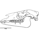
Ostéologie de la tête de Richardus excavans Lavocat,1988
Published online: 30/10/1989
Keywords:
Africa; anatomy; Bathyergidae; Miocene; Rodents
https://doi.org/10.18563/pv.19.2.73-80
Abstract
Remarkable association of a small infraorbital foramen, similar to that in recent Heterocephalus, and of a strong muscular print on the dorsal anterior part of the zygomatic plate and on the premaxillary. Several anatomical structures to be compared with those of Heterocephalus suggest relationships with this genus. Richardus supports the ancestrality of the hystricomorph character in the bathyergids
PV article infos
Published in Vol. 19, Fasc. 2 (1989)
|
PDF
|
