
Pterosaurs from Coahuila
Abstract book of the 18th Conference of the EAVP
Two enigmatic rodents from Lavergne (MP 16), Quercy Phosphorites
Les sélaciens du Miocène de la région de Montpellier
Muridae du Pliocène supérieur d'Espagne et du midi de la France.
Les Chiroptères du Miocène inférieur de Bouzigues. 1- Etude systématique.
Eocene (57) , Quercy Phosphorites (38) , Systematics (32) , Rodents (29) , Mammalia (27) , Rodentia (25) , Miocene (24)

|
Les mammifères Montiens de Hainin (Paléocène moyen de Belgique) Part III : MarsupiauxJean-Yves Crochet and Bernard SigéPublished online: 30/09/1983Keywords: Belgium; Marsupials; Paleobiogeography; Paleocene https://doi.org/10.18563/pv.13.3.51-64 Abstract The oldest european marsupials are described from some specimens (isolated upper molars) recently found from the Hainin sediment (Middle Paleocene of Belgium). These fossils document a new species of the Peradectes genus. They give evidence of a much older occurrence of the marsupials in Europe than it was assumed. They allow us to postulate a didelphid dispersal from South America towards the western-holarctic area operating in two phases : the first one of the Peradectes genus at the end of the Cretaceous; the second one of the Didelphíni tribe at the end of the Paleocene. A central american crossing is likely for the first one, whereas a transafrican way is tentatively argued for the second one. PV article infos Published in Vol. 13, Fasc. 3 (1983) |
|

|
Contributions à l'étude du gisement Miocène supérieur de Montredon (Hérault). Les grands mammifères. 4 - Les artiodactyles Suidae.Léonard GinsburgPublished online: 15/11/1988Keywords: Artiodactyla; France; Mammalia; Montredon; Upper Miocene https://doi.org/10.18563/pv.18.ext.57-64 Abstract There is only one suid known in the Upper Miocene of Montredon (Hérault): Microstonyx (Limnostonyx nov. subgen.) antiquus (KAUP). It is differenciated from Microstonyx major by the presence of upper and lower canines which are considerably longer and biger. Its presence at Montredon corroborates the palustrine habitat for the species. PV article infos Published in Vol. 18, Ext (1988) |
|

|
Les rongeurs du Miocène moyen et supérieur du MaghrebJean-Jacques Jaeger
Published online: 15/05/1977 |
|
|
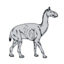
Reconstruction of the cervical skeleton posture of the recently-extinct litoptern mammal Macrauchenia patachonica Owen, 1838R. E. Blanco, Lara Yorio
Published online: 02/05/2023 |
|

|
Les Affinités de Nyctereutes megamastoides (Pomel), canidé du gisement Villafranchien de Saint-Vallier (Drôme, France).R. MartinPublished online: 31/01/1971Keywords: Canidae; Nyctereutes; Villafranchian https://doi.org/10.18563/pv.4.2.39-58 Abstract Nyctereutes megamastoides (Pomel) from the Villafranchian of the Auvergne and from Saint-Vallier presents cranial and dental characters sufficiently close to those of the late Pliocene canid from Perpignan (Roussillon), described by Depéret under the specific name of Canis donnezani belonging to the same genus Nyctereutes. The extinction of the European "Nyctereutes" group seems due to the too great alimentary specialization of this canid, whereas the Asiatic lineage represented in the Villafranchian by Nyctereutes sinensis Schlosser and at present by Nyctereutes procyonider Gray was able to maintain itself probably by means of a profound change in its alimentary regime. PV article infos Published in Vol. 04, Fasc. 2 (1971) |
|
|
Batoids (Rajiformes, Torpediniformes, Myliobatiformes) from the Sülstorf Beds (Chattian, Late Oligocene) of Mecklenburg, northeastern Germany: a revision and description of three new speciesThomas ReineckePublished online: 24/06/2015Keywords: Batoids; Chattian; Elasmobranchii; North Sea Basin; Oligocene https://doi.org/10.18563/pv.39.2.e2 Abstract Bulk-sampling of fossil-rich tempestites from the Chattian Sülstorf Beds of PV article infos Published in Vol.39-2 (2015) |
|
|
New Late Miocene plecotine bats (Chiroptera, Vespertilionidae: Plecotini) from Gritsev, UkraineValentina V. Rosina, Sergei Kruskop
Published online: 07/03/2019 |
|
|
A partial skeleton of Metaxytherium medium from the middle Miocene of La Morfassière quarry (Indre-et-Loire, France)Stéphane Ducrocq
Published online: 16/01/2025 |
|

|
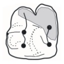
Nouvelle quantification de l'Hypsodontie chez les Theridomyidae : l'exemple de Theridomys ludensis nov. sp.Monique Vianey-Liaud
Published online: 30/12/1985 |
|

|
Observations sur des remaniements structuraux post-mortem dans des dents de mammifères fossiles provenant des phosphorites du QuercyJean-Albert RemyPublished online: 01/12/1974Keywords: Quercy Phosphorites; rearrangements; Teeth https://doi.org/10.18563/pv.6.3-4.163-176 Abstract Deux types de remaniements post mortem me paraissent caractéristiques de l'état de conservation des dents de mammifères fossiles dans les Phosphorites du Quercy : PV article infos Published in Vol. 06, Fasc. 3-4 (1975) |
|

|
Eggshell microstructure and porosity of the Nicobar scrubfowl (Megapodius Nicobariensis, great Nicobar island, India)Géraldine Garcia
Published online: 16/12/2008 |
|
|
A late Eocene palaeoamasiine embrithopod (Mammalia, Afrotheria) from the Adriatic realm (Island of Rab, Croatia)Fabrice Lihoreau
Published online: 14/12/2023 |
|

|
Les Bovidae (Artiodactyla, Mammalia) du Miocène moyen de la formation Hofuf (Province du Hasa, Arabie Saoudite).Herbert ThomasPublished online: 30/12/1983Keywords: Biostratigraphy; Bovidae; Middle Miocene; Palaeogeography; Saudi Arabia https://doi.org/10.18563/pv.13.5.157-206 Abstract The study of the bovids from Al Jadidah (Hofuf Formation, Saudi Arabia) confirms that the fauna comes from a pre-Hipparion level. The Al Jadidah age is close to that of Fort Ternan (14 m.y.) and Beni Mellal, but cannot be older than that of Fort Ternan. The age of the Hofuf Formation is close to but slightly older than the oldest deposits of the Ngorora Formation (Kenya). 7 to 9 species have been recorded, of which 2 to 4 remain indeterminate. If the great specific diversity of te bovids from this locality gives evidence of immigrations from anterior Asia (Turkey) (e.g. Pachytragus Iigabuei sp. nov.), the bovid assemblage of Al Jadidah results in fact from a double influence: from the anterior Asia and mainly from Africa (e.g. the Caprotragoides lineage and the Neotragini? Homoiodorcas). The Al Jadidah bovids reflect, on the whole, the predominant character of open to very open environment, which supports the conclusions drawn from our two preliminary studies. PV article infos Published in Vol. 13, Fasc. 5 (1983) |
|

|
Study of the Turolian hipparions of the lower Axios valley (Macedonia, Greece). 4. Localities of Dytiko.George D. KoufosPublished online: 15/12/1988Keywords: Equidae; Greece; Hipparion; Lower Axios Valley; Macedonia; Mammalia; Turolian https://doi.org/10.18563/pv.18.4.187-239 Abstract The hipparions from the Dytiko localities of the lower Axios valley (Macedonia, Greece) are studied. The material comes from three localities Dytiko-l, 2, 3 (DTK, DIT, DKO), which are situated near the village of Dytiko, about 60 km northwest to Thessaloniki. Three species have been determined, the medium-sized H. mediterraneum, the small-sized H. matthewi and the very small-sized H. periafricanum. The determined Hipparion species, their morphological characters and their comparison with the other Axios valley material indicate a Late Turolian age for the Dytiko localities. PV article infos Published in Vol. 18, Fasc. 4 (1988) |
|

|
Eléments nouveaux sur l'évolution des genres Eucricetodon et Pseudocricetodon (Eucricetodontinae, Rodentia,Mammalia, de l'Oligocène d'Europe Occidentale.Bernard ComtePublished online: 01/07/1985Keywords: evolution; Occidental Europe; Oligocene; Rodentia; Systematics https://doi.org/10.18563/pv.15.1.1-68 Abstract The review of material recently collected in the new localities from the “Phosphorites du Quercy", and different localities from the South of France, bring new informations on the genus Eucricetodon THALER. 1966, and Pseudocricetodon THALER, I969 (Middle and Upper Oligocene. Western Europe). Thanks to Eucricetodon huerzeleri VIANEY-LIAUD, 1972, which were unsufficiently known until now, is proposed. During the middle Oligocene Eucricetodon atavus MISONNE, 1957 seems to give rise to two lineages. One of them led to Eucricetodon huberi,which however exhibits a larger size and a development of progressive characters on the teeth. The other would be Eucricetodon huerzeleri well differentiated at the “Mas de Pauffié" standard level (beginning of the upper Oligocene). The ornementation of lower incisors is described, when possible. Though the fossils are not abundant, it seems that the ancestral lineage, Eucriretodon atavus, remains in the upper Oligocene (Boningen standard level). evolving into Eucricetodon praecursor SCHAUB, 1925 (Rickenbach standard level). The characters of Eucricetodon dubius (SCHAUB. 1925), represented by a numerous population in Pech Desse and Pech du Fraysse (Quercy). confirm that this species and Eucricetodon praecursor belong to two different lineages. As Eucricetodon dubius shows more primitive features, this species could not originale from Eucricetodon atavus -Eucricetodon huberi. The appearance of this species at the level of Mas de Pauffié could be the result of an immigration. A new definition of Pseudocricetodon incertus (SCHLOSSER. 1884) is given. This species has been found in several localities, where it had not been identified until now. lts comparison with Pseudocricetodon moguntiacus (BAHLO. l975), found at several localities from the standard level of Antoingt (end of middle Oligocene). shows a parallel evolution to that of Pseudocetodon incertus, which is of larger size and with a less complicated pattern of teeth. PV article infos Published in Vol. 15, Fasc. 1 (1985) |
|
|
A reassessment of the giant birds Liornis floweri Ameghino, 1895 and Callornis giganteus Ameghino, 1895, from the Santacrucian (late Early Miocene) of Argentina.Eric Buffetaut
Published online: 13/12/2016 |
|
|
New datation of the Tafna Basin (Algeria): A combination between biochronological and magnetostratigraphical dataSalamet Mahboubi
Published online: 11/03/2015 |
|

|
Rongeurs du Miocène inférieur et moyen en Languedoc. Leur apport pour les correlations Marin-Continental et la Stratigraphie.Jean-Pierre AguilarPublished online: 31/03/1980Keywords: Languedoc; Miocene; Rodents; Southern France https://doi.org/10.18563/pv.9.6.155-203 Abstract The rodents (Cricetidae, Gliridae, Sciuridae) found in lacustrine, brackish marine and karstic sediments of Miocene age in Languedoc, assign the position of the different localities in the scale of "niveaux repères" used by mammalogists. Some detailed stratigraphical studies bring several correlations between this continental biochronological scale and the marine scale ; the most important results are the Aquitanian age of the "niveaux repères" of Coderet and Paulhiac, the Burdigalian age of Laugnac, Estrepouy, Vieux-Collonges, La Romieu and Sansan and the Langhian or Lower Serravallian age of La Grive M. The correlations between the Tethys and the Central Paratethys for the Lower Neogene profit also of these results, since the locality of Neudorf Spalte 1, 2 (Czechoslovakia) is shown to be younger than Sansan (France). The paleontological study has also several geological inferences for the Miocene of Languedoc ; with the calibration of this Miocene, we know quite precisely that the Lower Miocene is chiefly a time lacustrine sedimentation, and also that the marine Miocene sedimentation ends early in the Miocene Period, in Langhian or lower Serravallian times. PV article infos Published in Vol. 09, Fasc. 6 (1980) |
|

|
Morphotypes dentaires actuels et fossiles des chiroptères vespertilionines. 2ème partie: implications systématique et phylogéniques.Henri MenuPublished online: 15/11/1987Keywords: Chiroptera; PHYLOGENY; Systematics; Vespertilionine https://doi.org/10.18563/pv.17.3.77-150 Abstract The first part of this study was devoted to a descriptive analysis of teeth morphologies among the vespertilionine bats. This leads now to a tentative synthesis, providing views on the systematics of the group. The results could be seen according to three distinct but closely related purposes : 1 - the sorting of the genera contents in order to conform the genera units to homogeneous taxa that could represent natural issues of evolutionary lineages ; 2 - the investigation of relationships between extant genera in order to infer the possibilities of common origin ; 3 - according to the preceeding items and to the observed evolutionary trends, a tentative phylogeny, modest and cautious. The contents of many genera are sorted : Leuconoe is removed from subgeneric to generic position, whereas Myotis becomes a subgenus of it ; the myotodont species are cleared away from the Pipistrellus genus ; Glischropus and Scotozous are synonymized within Pipistrellus ; Hypsugo is raised to the generic level ; some species previously ranged within Pipistrellus will form provisionally a collective group, Attalepharca nov. ; the Eptesicus genus is broken up, the excluded species being grouped within Nycterikaupius gen. nov. ; the Nycticeini tribe is defined again after exclusion of Otonycteris , Scotoecus, Scotophilus , and addition of Hesperoptenus ; the species la io and Pipistrellus tasmaniensis are removed to Eptesicus (n.s.) and Pipistrellus dormeri to Scotoecus. Groupings of genera are stated according to the main evolutionary trends of I1/. The relevance of these is often warranted by close morphologic similarities of other teeth. This leads to a recognition of the major evolutionary radiations which occurred in the group. The filiations schematized at the end of the work show the dental relationships observed between the extant genera, and could represent a phylogenic framework. Two major facts are to be underlined : 1- the early divergence of leuconoids ; 2 - the successives crossings to myotodonty from the nyctaloid flow. Fossil data from the literature are punctually and tentatively incorporated within phylogenic sketches. PV article infos Published in Vol. 17, Fasc. 3 (1987) |
|

|
Contribution à l'étude des genres Gliravus et Microparamys (Rodentia) de l'Eocène d'Europe.Jean-Louis HartenbergerPublished online: 15/03/1971Keywords: Eocene; Gliravus; Microparamys; Rodentia https://doi.org/10.18563/pv.4.4.97-135 Abstract Based on material found in about 15 localities the relationships of the genera Microparamys and Glirarus have been studied. One new genus, two subgenera and three species [Microparamys (Sparnacomys) chandoni n. subgen. and n. sp., Microparamys (Pantrogna) russelli n. subgen., Eoglirarus wildi n. gen. and n. sp., Gliravus meridionalis n. sp.] as well as the publication PV article infos Published in Vol. 04, Fasc. 4 (1971) |
|