
Pterosaurs from Coahuila
Abstract book of the 18th Conference of the EAVP
Two enigmatic rodents from Lavergne (MP 16), Quercy Phosphorites
Les sélaciens du Miocène de la région de Montpellier
Muridae du Pliocène supérieur d'Espagne et du midi de la France.
Les Chiroptères du Miocène inférieur de Bouzigues. 1- Etude systématique.
Eocene (57) , Quercy Phosphorites (38) , Systematics (32) , Rodents (29) , Mammalia (27) , Rodentia (25) , Miocene (24)
|
Eocene Teleostean Otoliths, Including a New Taxon, from the Clinchfield Formation (Bartonian) in Georgia, USA, with Biostratigraphic, Biogeographic,
|
|

|
Sur le statut taxonomique de Myotis KAUP 1829 (Mammalia, Chiroptera).Henri MenuPublished online: 15/12/1988Keywords: Myotis; taxonomy https://doi.org/10.18563/pv.18.4.263 Abstract Suite à la récente publication d'une révision systématique des Chiroptera Vespertilioninae, conduite sur la base des morphologies dentaires comparées (Menu 1987), une remarque bienveillante du Dr. V. Aellen (Muséum d'Histoire Naturelle de Genève) a attiré l'attention de l'auteur sur un point précis du Code International de Nomenclature Zoologique. PV article infos Published in Vol. 18, Fasc. 4 (1988) |
|

|
Préface au mémoire jubilaire en hommage à René LavocatJacques MichauxPublished online: 01/10/1980Keywords: Editorial https://doi.org/10.18563/pv.9.ext.1-13 Abstract Monsieur René Lavocat, Directeur du Laboratoire de Paléontologie des Vertébrés de la troisième section de l'Ecole Pratique des Hautes Etudes, quittait le service actif en l'année 1979. View editorial Published in Vol. 9, Ext (1980) |
|

|
Révision des Chiroptères Lutériens de Messel (Hesse, Allemagne).Donald E. Russell and Bernard SigéPublished online: 04/04/1970Keywords: Chiroptera; Lutetian; Messel https://doi.org/10.18563/pv.3.4.83-182 Abstract The revision of the Lutetian chiropterans from Messel, first described by Revilliod in 1917, is based on the anatomy of the teeth and the skeleton. A figuration or refiguration of thematerial utilized accompanies the new description, which goes beyond that of the original monograph. PV article infos Published in Vol. 03, Fasc. 4 (1970) |
|

|
Les vertébrés fossiles de Colombie et les problèmes posés par l'isolement du Continent sud-Américain.Jaime de PortaPublished online: 20/01/1969Keywords: Columbia; Cretaceous; Fauna; Quaternary; South America https://doi.org/10.18563/pv.2.2.77-94 Abstract A general view is given of the vertebrate faunas, Cretaceous to Quaternary of age, found in Columbia and of their principal characteristics. This view leads to the discussion of the isolation of the South American continent and of the role played by the Bolivar syncline with respect to North American immigrants during the Oligocene. The absence of marine deposits of Oligocene age in the north and northwest of Columbia suggests the possibility of a communication with Central America. This communication would have permitted the passage of hystricomorph rodents, of platyrrhine monkeys, and of colubrids. The non-occupation, until then, of the ecologie niches of these groups would have favored their installation beside the indigenous fauna. In this hypothesis it would no longer be necessary to admit that these vertebrates arrived as «island hoppers ››. The eco-biologic conditions would explain the absence of large-sized forms of North American origin. PV article infos Published in Vol. 02, Fasc. 2 (1969) |
|

|
Les rongeurs du site Pliocène à Hominidés de Hadar (Ethiope)Maurice SabatierPublished online: 15/02/1982Keywords: Ethiopia; hominids; Muridae; Pliocene https://doi.org/10.18563/pv.12.1.1-56 Abstract The intensive exploration of the Pliocene Hadar Formation, rich in hominid remains, led us to the discovery of several micromammals levels. ln some of them, rodents are very abundant. The stratigraphic repartition of these levels do not cover the whole fossiliferous series of the formation but takes place only in the sedimentary members from Sidi Hakoma and Denen-Dora (rancing from 3.1 - 3.2 MY to 2.8 - 2.9 MY, according to the recent geochronological data). During this gap of time, the species do not show morphological changes, what allowed us to gather, in the same taxa, forms of slighty different ages. PV article infos Published in Vol. 12, Fasc. 1 (1982) |
|

|
Pantolestidae nouveaux (Mammalia, Insectivora) de l'Eocène moyen de Bouxwiller (Alsace).Jean-Jacques Jaeger
Published online: 31/03/1970 |
|
|
Book of Abstracts of the XXII Annual Meeting of the European Association of Vertebrate Palaeontologists, 30 June–5 July 2025, Kraków, PolandGeorgios L. Georgalis
Published online: 20/06/2025 |
|

|
Contributions à l'étude du gisement miocène supérieur de Montredon (Hérault). Les grands mammifères. Avant propos.Bernard SigéPublished online: 15/11/1988Keywords: Editorial; Mammalia; Montredon; Upper Miocene https://doi.org/10.18563/pv.18.ext.1-2 Abstract Le Mémoire Extraordinaire 1988 de PALAEOVERTEBRATA regroupe dix articles consacrés au gisement à mammifères du Miocène supérieur de Montredon (Hérault), connu et classique depuis la fin du siècle dernier, et auquel est lié le nom du savant paléontologue lyonnais Charles Depéret. View editorial Published in Vol. 18, Ext (1988) |
|

|
Les Amphibiens et les reptiles du Pliocène supérieur de Balaruc II (Herault, France)Salvador Bailon
Published online: 15/09/1989 |
|
|
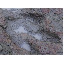
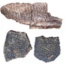
Late Campanian theropod trackways from Porvenir de Jalpa, Coahuila, MexicoHéctor E. Rivera-Sylva
Published online: 29/11/2017 |
|
|
Les rongeurs de Chéry-Chartreuve et Rocourt-Saint-Martin (est du bassin de Paris; Aisne, France). Leur place parmi les faunes de l'Eocène Moyen d'EuropeBernard Comte, Maurice Sabatier and Monique Vianey-Liaud
Published online: 15/12/2012 |
|

|
Contributions à l'étude du gisement Miocène supérieur de Montredon (Hérault). Les grands mammifères. 7 - Les proboscidiens DeinotheriidaeHeinz TobienPublished online: 15/11/1988Keywords: allometry; Astaracian; Deinotherium; Montredon; Systematics; taphonomy; Vallesian https://doi.org/10.18563/pv.18.ext.135-175 Abstract Some complete tooth rows and about one hundred isolated teeth enabled the identification of the deinothere of the Vallesian site Montredon (Hérault) as Deinotherium giganteum KAUP 1829, mainly by comparisons with the likewise Vallesian sample of the type locality Eppelsheim (Rheinhessen, F.R.G.). PV article infos Published in Vol. 18, Ext (1988) |
|

|
Ein neuer condylarthre und ein tillodontier (Mammalia) aus dem Mitteleozän des Geiseltales.Jens L. Franzen and Hartmut HauboldPublished online: 15/04/1986Keywords: Condylarthra; Eocene; Europe; Mammalia; taxonomy; Tillodontia https://doi.org/10.18563/pv.16.1.35-53 Abstract In the course of a revision of the Equoidea numerous dentitions as well as a partial skeleton of a Phenaeodont were discovered from the Middle Eocene lignite beds of the Geiseltal locality. These fossils are recognized as a new genus and species of Phenacodontidae : HaIlensia matthesi n.g. n.sp.. The species is present in the « untere und obere Unterkohle ›› (uUK, oUK = the lower and upper part of the Lower Coal Seam) as well as in the « obere Mittelkohle ›› (oMK = the upper part of the Middle Coal Seam). Two fragmentary upper jaws described and figured by Matthes (1977) as Propachynolophus gaudryi are also belonging to Hallensia matthesi. Thus the decisive argument for classifying the " Unterkohle " of the Geiseltal section as Lower Eocene has to be dropped. Another relict form of the Geiseltal is Esthonyx tardus n. sp. documented by a fragmentary mandible coming from the « untere Unterkohle ››. This is the latest Tillodont from Europe. Contrasting to E. munieri from the european Lower Eocene the dentition of E. tardus is morphologically more progressive. PV article infos Published in Vol. 16, Fasc. 1 (1986) |
|

|
Macroscelidea, Insectivora and Chiroptera from the Miocene of east Africa.Percy M. ButlerPublished online: 15/11/1984Keywords: Chiroptera; East Africa; Insectivora; Macroscelidea; Miocene; Systematics https://doi.org/10.18563/pv.14.3.117-198 Abstract The East African Miocene Macroscelidea, lnsectivora and Chiroptera are revised on the basis of new material. New taxa proposed are: Miorhynchocyon, .n. gen. (Macroscelididae): Míorhynchocyon meswae, n. sp.: Pronasílío ternanensis. n. gen.. n. sp. (Macroscelididae); Hiwegicyon juvenalis, n. gen. n. sp. (Macroscelididae); Parageogale, n. gen. (Tenrecidae): Prochrysochlorinae, n. subfam. (Chrysochloridae): Propottininae, n. subfam, (Pteropodidae); Chamtwaria pickfordi, n. gen., n. sp. (Vespertilionidae). Gymnurechnínus songhorensis is synonymised with G. camptolophus. The new material provides additional information on the dentition, especially of Myohyrax oswaldi. Galerix africanus. Amphechínus rusingensis, Protenrec tricuspis and Parageogale aletris. Partial skulls are described of Amphechinus rusingensis, Protenrec tricuspis, Prochrysochloris míocaenicus and Taphozous incognita. The oldest member of the Macroscelidinae (Pronasilio) is described from Fort Ternan. Galerix africanus is closely related to G. exilis from Europe. Amphechinus rusingenesis is compared with Asiatic Oligocene Erinaceinae. The Miocene age of Crocidura is rejected. On the evidence of humeri, the following families of Chiroptera are newly reported: Pteropodidae, Nycterididae, Vespertilionidae, Molossidae. Propotto is regarded as an offshoot from the Pteropodidae, not ancestral to modern forms. Chamtwaria is a primitive vespertilionoid, provisionally placed in the Kerivoulinae. Erinaceidae probably entered Africa at the beginning of the Miocene, before 20 Ma. Faunistic differences between deposits are largely to be ascribed to differences in local environment. PV article infos Published in Vol. 14, Fasc. 3 (1984) |
|

|
Die Ohr-Region der Paulchoffatiidae (Multituberculata, Ober-Jura).Gerhard HahnPublished online: 15/11/1988Keywords: Multituberculata; Ober-Jura; Paulchoffatiidae; Petrosum; Portugal https://doi.org/10.18563/pv.18.3.155-185 Abstract The petrosal of the Paulchoffatiidae HAHN, 1969 is described and compared with that of younger multituberculates and of other Mesozoic mammals. The "Morrison petrosal", described by Prothero (1983), is also discussed; it probably belongs to the multituberculates. The reconstruction of the ventral side of the Paulchoffatiinae-skull, given by Hahn in 1987, is completed by addition of the otic and the occipital region. PV article infos Published in Vol. 18, Fasc. 3 (1988) |
|

|
Les Gliridés (Rodentia) de l'Oligocène supérieur de Saint-Victor-la-Coste (Gard).Marguerite HugueneyPublished online: 28/10/1968Keywords: Gliridae; Late Oligocene https://doi.org/10.18563/pv.2.1.1-16 Abstract The locality of St.-Victor-la-Coste (Gard) has yielded, rather abundantly, teeth of two glirids hitherto very poorly known: Glirudinus praemurinus (Freudenberg) and Glirudinus glirulus (DEHM). It has permitted, moreover, new views on the evolution of Peridyromys murinus (POMEL). Study of these forms confirms the late Oligocene age of the fauna, without allowing, however, further precision. PV article infos Published in Vol. 02, Fasc. 1 (1968) |
|

|
Insectivores pliocènes du Sud de la France (Languedoc-Roussillon) et du Nord-Est de l'Espagne.Jean-Yves CrochetPublished online: 31/10/1986Keywords: Biostratigraphy; Insectivora; Languedoc; Pliocene; Spain; Systematics https://doi.org/10.18563/pv.16.3.145-171 Abstract The first lists of Insectivores (Erinaceidae, Talpidae and Soricidae) from the Pliocene beds of Southern France and North-East Spain are given in this paper. The material from twelve localities is studied. These localities are geographically situated in Languedoc (Celleneuve, Vendargues, Nîmes, Sète, Balaruc 2 and Seynes), in Roussillon (Terrats, Serrats-d'en-Vacquer, Château d'eau and Mont-Hélène) and in North-East Spain (Layna, Medas Islands and Puebla de Valverde). These faunas correspond to the Early, Middle and Late Pliocene. 1 to 8 taxa are identified in these localities and 14 specific taxa are presently listed for this period in this area. Two new specific taxa are described as Galerix depereti nov. sp. from all the Early Pliocene localities in the North-Pyrenean area and as Desmanella gardiolensis nov. sp. from Balaruc 2. For this small mammals, two faunal assemblages are recognized. The first one is dated from the Early Pliocene (F 1, 2 and 3 zones in Aguilar et Michaux) and is characterized by Galerix depereti and rare and little diversified Soricids. The second one is Late Pliocene in age (zones G 2 and G 3). The fossils of the genus Talpa are relatively abundant and the Soricids are diversified and very abundant. The Middle Pliocene (zone G 1) is a transitional period. ln these faunas, most of the insectivore genera are known from the European Late Miocene beds (8 on 10). This fact demonstrates a relative continuity between the invectivore faunas from the Late Miocene to the Early Pliocene. In conclusion, somme paleoecological considerations are suggested. PV article infos Published in Vol. 16, Fasc. 3 (1986) |
|
|
Terrestrial vertebrate paleocommunities from the Cerro del Pueblo Formation (Late Cretaceous; Late Campanian) at Las Aguilas, Coahuila, MexicoHéctor E. Rivera-Sylva
Published online: 16/07/2019 |
|

|
Neue Beobachtungen zum Schädel-und Gebiss-Bau der Paulchoffatiidae (Multituberculata,Ober-Jura).Gerhard HahnPublished online: 15/12/1987Keywords: Dentition; Paulchoffatiidae; Portugal; Skull structure; Upper Jurassic https://doi.org/10.18563/pv.17.4.155-196 Abstract The ventral face of the Paulchoffatiinae skull (Multituberculata, Lower Kimmeridgian, Portugal) is new reconstructed. Some details hitherto unknown are added, as the presence of jugals, the structure of the palatine and the extension of the pterygoids. The situation of the m2/ is discussed. Kielanodon hopsoni n. g., n. sp. is erected, known by its upper p3-5/. From Guimarotodon leiriensis the mandible with its dentition is made known. New informations concerning the milk-dentition and the replacement of teeth are also added. PV article infos Published in Vol. 17, Fasc. 4 (1987) |
|