
|
This review critiques the third edition (1986) of The Animals (Systematische Zoologie), a descriptive zoology textbook first published in 1976. The book focuses on morphology, taxonomy, and basic anatomy of animal groups, with occasional notes on distribution, habits, and economic/medical significance.
Published online: 15/04/1986
Keywords:
Book review; Systematics
https://doi.org/10.18563/pv.16.1.55
Abstract
THE ANIMALS. Systematische Zoologie by A. REMANE, V. STORCH, and U. WELSCH. 1986, Edition 3. Cloth and paperbound. Stuttgart and New York : Gustav Fischer Verlag. xvi + 698 pp. DM 86. (Geb.) & DM 72 (kart.).
PV article infos
Published in Vol. 16, Fasc. 1 (1986)
|
PDF
|

|
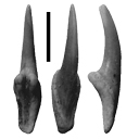
Types dentaires adaptatifs chez les sélaciens actuels et post-paléozoïques.
Published online: 01/09/1986
Keywords:
Dental types; evolution; Fossil selachians; Recent selachians; Trophic adaptations
https://doi.org/10.18563/pv.16.2.57-76
Abstract
The dentition of selachians is characterized by an often very pronounced heterodonty involving a great morphological diversity. Despite this fact, the dentitions of selachians can be grouped in a rather reduced number of dental types corresponding to trophic adaptations: grasping, tearing, cutting, crushing, grinding and grasping-grinding type. The numerous exemples of convergence and parallelism that can be observed in fossil selachians and between Recent and fossil ones is the result of this reduced number of dental types. These dental specialisations allow to try a reconstruction of the way of life of fossil forms.
PV article infos
Published in Vol. 16, Fasc. 2 (1986)
|
PDF
|

|
Pairomys et Ectropomys : la fin d'une ambiguïté ; mise au point sur les Oltinomyinae et Remyinae (Rodenia, Theridomyidae).
Published online: 20/05/1994
Keywords:
PHYLOGENY; taxonomy; Theridomyidae; Upper Eocene
Abstract
The description of new material of the Upper Eoœne of France, Southem Germany and Spain together with the restudy of the material described by Thaler (1966b) as Pairomys allows to confirm the validity of the genus Ectropomys up to know controversally discussed. As a consequence the subfamily Remyinae and Oltinomyinae can be defined precisely. The study of the dental characters shows that the Remyinae in fact belong to the Theridomyidae and represent a derived evolutionary unit within this family. The Oltinomyinae together with the Theridomyinae and Issiodoromyinae very probably form a monophyletic unit.
PV article infos
Published in Vol. 23, Fasc. 1-4 (1994)
|
PDF
|

|
Une faunule de vertébrés sous la base de grès de Celas (Eocène supérieur) à ST Dresery (Gard)
Published online: 20/05/1994
Keywords:
Artiodactyla; Biostratigraphy; Eocene; Mammals
Abstract
The St-Dézéry local fauna (3 reptile-, 4 mammal species) is approximately of the same age as the La Débruge or the Ste-Néboule faunas. It conduces to a better dating of the limestones underlying the Célas sandstones. A large part of a mandible of Amphimeryx was found there, which documents the record of this family of small artiodactyls
PV article infos
Published in Vol. 23, Fasc. 1-4 (1994)
|
PDF
|

|
Mammifères de l'Ilerdien Moyen (Eocène inférieur) des Corbières et du Minervois (Bas-Languedoc, France). Systématique, Biostratigraphie, Corrélations.
Published online: 25/01/1991
Keywords:
Biostratigraphy; Corbières; correlations; Early Eocene; Ilerdian; Mammalia; Minervois; Paleobiogeography; Southern France
Abstract
Mammal-bearing localities have been discovered in the marine and lacustrine series of the middle Ilerdian (Lowermost Eocene) from Southem France (Minervois and Corbières). In the localities of Fordones, Monze, Fournès, and La Gasque, thirty mammal species have been identified. Among others, they include ischyromyid rodents (Microparamys and Pseudoparamys), paromomyid and adapid primates (Arcius and Donrussellia), new insectivores, condylarths, and a dyspternine pantolestid. These faunas provide new informations on the early Eocene Mesogean faunas of Rians and Palette. The assemblages of primates and rodents from Fordones support good correlations with Palette which was recently placed near the standard-level of Dormaal (MP 7). In fact, Palette and Fordones could be even older than Dormaal. Consequently, there seems to be a relatively important temporal gap between the late Paleocene of Cernay and the Sparnacian of Dormaal. This gap could be partly filled with the Mesogean faunas of Palette, Fordones, and Silveirinha. On the basis of these new mammal faunas the marine middle Ilerdian is proved to be older than the Cuisian stage of the Paris Basin. With regards to the position of the Fordones fauna at the top of the NP 10 calcareous nannoplankton biozone, the westem European paleomammalogists Paleocene/Eocene boundary could be situated between the NP 9 and NP 10 biozones.
PV article infos
Published in Vol. 20, Fasc. 2-3 (1991)
|
PDF
|

|
Editorial
Published online: 29/04/1991
Keywords:
Editorial
Abstract
Editorial for celebrating the publication of the volume 20.
La revue PALEOVERTEBRATA a été fondée en 1967 par Louis THALER, à une époque où il n'existait, au plan international, que deux périodiques uniquement consacrés aux vertébrés fossiles. Actuellement ce nombre n'est que de quatre. A l'origine la revue fut essentiellement créée pour diffuser les résultats des chercheurs du Laboratoire de Paléontologie de l'Université de Montpellier, alors en début d'expansion et plusieurs thèses et de nombreux travaux directement liés à des thèses y furent publiés ce qui n'excluait déjà pas la publication d'articles d'auteurs étrangers.
[...]
View editorial
Published in Vol. 20, Fasc. 4 (1991)
|
PDF
|

|
Nouveau Dichobunidae (Artiodactyla, Mammalia) du gisement d'Aumelas (Hérault) d'âge Lutétien terminal
Published online: 01/10/1980
Keywords:
Aumelas; Dichobunidae; Hérault; Middle Eocene; Upper Lutetian
https://doi.org/10.18563/pv.9.ext.197-211
Abstract
The faunal list of the mammals collected at the locality of Aumelas (Hérault, France) is revised. For the first
time this Middle Eocene locality is precisely settled in the european chronological scale of "niveaux repères", between the levels of Bouxwiller and Egerkingen, in Uppermost Lutetian.
A new Dichobunid from the site is described : Aumelasia gabineaudi n. g., n. sp. This new genus has primitive characters. and it may be in the descent of the Lower Eocene Protodichobune.
PV article infos
Published in Vol. 9, Ext (1980)
|
PDF
|

|
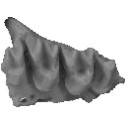
Cricetid rodents from Siwalik deposits near Chinji village. Part I: Megacricetodontinae, Myocricetodontinae and Dendromurinae.
Published online: 15/10/1988
Keywords:
Dendromurinae; Megacricetodontinae; Middle Miocene; Myocricetodontinae; Rodents; Siwalik
https://doi.org/10.18563/pv.18.2.95-154
Abstract
Seventeen species of cricetid rodent are recognized and described from lower and middle Siwalik deposits in the Potwar Plateau of Pakistan. These species are grouped in three categories, characterized as subfamilies (e. g., Megacricetodontinae, Myocricetodontinae, and Dendromurinae); an additional and more abundant category of rodents from these deposits, the Democricetodontinae, is excluded from this study, and will be described in a later study. Fifteen of the species are new, and four new genera are described. The Siwalik cricetid taxa are : Megacricetodon aquilari, n. sp.; Megacricetodon sivalensis, n. sp.; Megacricetodon daamsi, n. sp.; Megacricetodon mythikos, n. sp.; Punjabemys downsi, n. gen. & n. sp.; Punjabemys leptos, n. gen. & n. sp.; Punjabemys mikros, n. gen. & n. sp.; Myocricetodon sivalensis, n. sp.; Myocricetodon sp.; Dakkamyoides lavocati, n. gen. & n. sp.; Dakkamyoides perplexus, n. gen. & n. sp.; Dakkamys asiaticus, n. sp.; Dakkamys barryi, n. sp.; Dakkamys sp.; Paradakkamys chinjiensis, n. gen. & n. sp.; Potwarmus primitivus, n. gen.; and Potwarmus minimus, n. gen. & n. sp. This diverse record of middle Miocene small mammals illuminates a profound radiation of cricetid rodents in southem Asia, the effects of which were felt in Europe and Africa as well as the rest of Asia.
PV article infos
Published in Vol. 18, Fasc. 2 (1988)
|
PDF
|

|
La poche à phosphate de Ste-Neboule (Lot) et sa faune de vertébres du Ludien Supérieur. 1 La poche et son remplissage
Published online: 15/09/1978
Keywords:
Eocene; Quercy Phosphorites
https://doi.org/10.18563/pv.8.2-4.171-173
Abstract
La poche de Ste-Néboule, commune de Béduer (Lot), 15 km environ à l'WSW de Figeac, fait partie du groupe le plus septentrional des gouffres creusés par les ruissellements du Paléogène dans les calcaires jurassiques de la bordure sud-ouest du Massif Central et qui furent comblés à la même époque par des argiles sidérolithiques accompagnées de phosphate de chaux concrétionné ainsi que des restes de la célèbre faune dite «des phosphorites du Quercy» .
PV article infos
Published in Vol. 08, Fasc. 2-4 (1978)
|
PDF
|

|
La poche à phosphate de Sainte-Néboule (Lot) et sa faune de vertébrés du Ludien supérieur. 13-Rongeurs
Published online: 25/09/1978
Keywords:
Eocene; Quercy Phosphorites
https://doi.org/10.18563/pv.8.2-4.313-318
Abstract
Sainte-Néboule has yielded only 4 species of Rodents. But the Theridomyids (Blainvillimys rotundidens and Patriotheridomys altus) are very significative of the age of the locality: Ste-Néboule is lower than the marker level of Escamps
PV article infos
Published in Vol. 08, Fasc. 2-4 (1978)
|
PDF
|
|
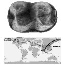
First early Eocene tapiroid from India and its implication for the paleobiogeographic origin of perissodactyls
Published online: 08/09/2015
Keywords:
Ceratomorpha; Helaletidae; Paléogène; Tapiromorpha; Vastan
https://doi.org/10.18563/pv.39.2.e5
Abstract
The presence of cambaytheres, the sister group of perissodactyls, in western India near or before the time of collision with Asia suggests that Perissodactyla may have originated on the Indian Plate during its final drift towards Asia. Herein we reinforce this hypothesis by reporting two teeth of the first early Eocene tapiromorph Perissodactyla from the Cambay Shale Formation of Vastan Lignite Mine (c. 54.5 Ma), Gujarat, western India, which we allocate to a new genus and species, Vastanolophus holbrooki. It presents plesiomorphic characters typical of the paraphyletic “Isectolophidae,” such as small size and weak lophodonty. However, the weaker hypoconulid and low paralophid, higher cusps, lower cristid obliqua, and the lingual opening of the talonid are found in Helaletidae, the most primitive tapiroid family. V. holbrooki, gen. et sp. nov., may be the oldest and the most primitive tapiroid, suggesting that at least tapiroid perissodactyls originated on India.
PV article infos
Published in Vol.39-2 (2015)
|
PDF
|
|
Autopsie d’une radiation adaptative : Phylogénie des Theridomorpha, rongeurs endémiques du Paléogène d’Europe - histoire, dynamique évolutive et intérêt biochronologique
Published online: 15/12/2016
Keywords:
Diversification; Extinction; Paléoenvironnements; Rodentia; Theridomyoidea
https://doi.org/10.18563/pv.40.3.e1
Abstract
Résumé :
L’étude des rongeurs Theridomorpha permet de suivre le déroulement d’une radiation adaptative pendant toute sa durée (Eocène moyen-Oligocène terminal), sur un territoire restreint à l’extrémité ouest de l’Europe Occidentale. Dans ce papier, la phylogénie de ce groupe est établie à partir d’une analyse cladistique reposant sur l’examen de 315 caractères (310 dentaires). Le groupe d’intérêt comprend 110 des 132 espèces (24 genres) de Theridomyoidea et deux genres encore inclus jusqu’ici dans les Reithroparamyinae qui rejoignent les Theridomorpha. Les groupes externes comprennent des Glires basaux, Cocomys, Tanquammys et 16 Ischyromyiformes. Un cadre phylogénétique robuste est produit, qui permet de clarifier la systématique des Theridomorpha. La position des Remyoidea (nov. sup.fam.) au sein des Ischyromyiformes, extérieure aux Theridomorpha, est confortée. Les Protadelomys et Tardenomys sont à la base des Theridomyoidea, avant la séparation en deux clades correspondant aux familles Pseudosciuridae et Theridomyidae. Les sous-familles sont consolidées : Pseudosciurinae et Sciuroidinae pour les Pseudosciuridae ; Issiodoromyinae, Oltinomyinae, Columbomyinae, Theridomyinae, auxquelles s’ajoute au moins une nouvelle sous-famille (Patriotheridomyinae), pour les Theridomyidae. La topologie des chrono-espèces (sensu Simpson), traitées antérieurement comme lignées évolutives, apparaît dans la plupart des cas sous forme de clades successifs dans lesquels les espèces sont le plus souvent arrangées de manière pectinée, émergeant dans l’ordre stratigraphique. L’analyse des caractères aux principaux nœuds permet de consolider les caractères diagnostiques des taxons et les tendances évolutives, ainsi que de discuter des divers parallélismes et convergences dans l’évolution des structures et patrons dentaires (e.g., émail des incisives unisérié chez les Issiodoromyinae et les Patriotheridomyinae, ou pseudo-multisérié chez les Blainvillimys les plus hypsodontes, les Protechimys et Archaeomys ; patrons dentaires téniodontes ; allongement des dents déciduales chez les Patriotheridomyinae, Issiodoromyinae et Theridomyidae ; sélénodontie ou lophodontie). Les dynamiques évolutives traduites par les changements morphologiques sont mises en relation avec les variations environnementales. Enfin, les implications biochronologiques de l’évolution des Theridomyoidea sont discutées.
Abstract:
The adaptive radiation of the rodents Theridomorpha occurred during a limited time window (middle Eocene to late Oligocene), on an area restricted to Western Europe. In this paper, the phylogeny of this group is established via a cladistic assessment of 315 morphological characters (310 dental). The group of interest encompasses 110 upon 132 species (24 genera) of Theridomyoidea, and two genera formerly included within the Reithroparamyinae, and which are included here within the Theridomorpha. The outgroups include basal Glires, Cocomys, Tanquammys and 16 Ischyromyiformes. A robust phylogenetic frame is produced, which allows clarifying the systematics of the Theridomorpha. Within the Ischyromyiformes, the Remyoidea (nov. supfam.) are set apart from the Theridomorpha. Protadelomys and Tardenomys represent the earliest offshoots of the Theridomyoidea, before the dichotomy between Pseudosciuridae and Theridomyidae. It supports the former subfamilies Pseudosciurinae and Sciuroidinae within the Pseudosciuridae; and for the Theridomyidae: the Issiodoromyinae, Oltinomyinae, Columbomyinae, Theridomyinae, with at least one new subfamily (Patriotheridomyinae). The topologies of the chronospecies (sensu Simpson), formerly considered as evolutionary lineages, appear in most cases as successive clades, in which the species are generally pectinately arranged and emerging in the stratigraphic order. The analysis of characters supporting the main nodes allow consolidating the diagnosic characters of the taxa and their evolutionary trends, as well as discussing the various cases of parallelism and convergence in the evolution of structures and dental patterns (e.g., uniserial incisor enamel for Issiodoromyinae and Patriotheridomyinae, or pseudo-multiserial for the most hypsodont Blainvillimys, Protechimys and Archaeomys; taeniodont dental patterns; lengthening of deciduous premolars for Patriotheridomyinae, Issiodoromyinae and Theridomyidae; selenodonty or lophodonty).
Evolutionary dynamics are analysed with respect to environmental changes. Finally, biochronological implications of the evolution of Theridomyoidea are discussed.
PV article infos
Published in Vol 40-3 (2016)
|
PDF
S.I. Data
|
|
A new species of hippopotamine (Cetartiodactyla, Hippopotamidae) from the late Miocene Baynunah Formation, Abu Dhabi, United Arab Emirates
Published online: 07/04/2017
Keywords:
Arab Peninsula; Hippopotamidae; Hippopotamine event; Systematics
https://doi.org/10.18563/pv.41.1.e2
Abstract
The discovery of new hippopotamid material from the late Miocene Baynunah Formation (Abu Dhabi, United Arab Emirates) has prompted the revision of the existing material of this as yet unnamed fossil taxon. The Baynunah hippopotamid appears to be distinct from all other contemporary and later species in having a relatively more elongate symphysis, a feature similar to the earlier (and more primitive) Kenyapotamus. Yet, the Baynunah hippopotamid presents a dentition typical of the Hippopotaminae. It is therefore a distinct species attributed to the later subfamily, described and named in this contribution. This species provides further evidence for a ca. 8 Ma evolutionary event (termed “Hippopotamine Event”) that initiated the spread and ecological significance of the Hippopotaminae into wet habitats across Africa and Eurasia. The morphological affinities of the new species from Abu Dhabi suggest that the Arabian Peninsula was not a dispersal route from Africa toward southern Asia for the Hippopotamidae at ca. 7.5 Ma to 6.5 Ma.
PV article infos
Published in Vol 41-1 (2018)
|
PDF
S.I. Data
|
|
A new species of bat (Chiroptera: Vespertilionidae) from the early Oligocene global cooling period, Brule Formation, North Dakota, USA
Published online: 09/12/2019
Keywords:
Eocene-Oligocene global cooling; Mammalia; Oligocene; Plecotini; Quinetia
https://doi.org/10.18563/pv.42.2.e2
Abstract
We report the first confirmed fossil bats from North Dakota, including a new species referable to the Vespertilionidae represented by a maxilla with P4-M3 from the Brule Formation, Fitterer Ranch local fauna, early Oligocene, Whitneyan North American Land Mammal Age. Unassociated postcranial fragments of the humerus and femur also represent a vespertilionoid, but appear to reflect a different, unidentified species. The new taxon, Quinetia frigidaria sp. nov., is referred to the genus Quinetia, previously known only from approximately contemporaneous deposits in Europe. The new species is larger than Quinetia misonnei from the early Oligocene of Belgium. It is similar in some morphological characters to Chadronycteris rabenae (Chiroptera incertae sedis) of the late Eocene (Chadronian) of northwestern Nebraska and to Stehlinia species (?Palaeochiropterygidae) from the Eocene and Oligocene of Europe, but differs from each in morphological details of the dentition and maxilla. An unassociated talonid of a lower molar from Fitterer Ranch shows myotodont morphology, unlike the nyctalodont lower molars in Q. misonnei, and thus represents a second chiropteran taxon in the fauna. Quinetia frigidaria is a member of a Paleogene radiation of bats near the low point of the Eocene-early Oligocene decline in global temperatures, increased seasonal aridity, and loss of tropical floras from mid-latitude North America.
PV article infos
Published in Vol 42-2 (2019)
|
PDF
|
|
Difficulties with the origin of dinosaurs: a comment on the current debate
Published online: 01/07/2020
Keywords:
dinosaur anatomy; dinosaur evolution; Ornithoscelida; palaeobiogeography; Triassic Period
https://doi.org/10.18563/pv.43.1.e3
Abstract
The origin and early evolutionary history of the dinosaurs is a topic that has recently gone through a period of renewed interest and academic debate. For 130 years, one way of classifying the various dinosaur subgroups persisted as the accepted model, with increasing levels of research in the past quarter-century also providing evidence for the hypothesis that dinosaur origination occurred in the Southern Hemisphere, particularly in South America. It is, after all, from within the Late Triassic strata of countries like Argentina and Brazil that we get some of the very best early dinosaur specimens; many of these specimens are the earliest known representatives of some of the major dinosaur subgroups, such as the theropods and sauropodomorphs. However, some recent analyses have brought about a shift in terms of what is currently accepted and what is now disputed regarding the origin of dinosaurs – the Southern Hemisphere origination hypothesis was questioned (although this was based upon observations and not with quantitative analysis techniques), as has the shape of the dinosaur tree. Responses to the new hypothesis were numerous; many further supported a Southern Hemisphere point of origin. Whilst the interrelationships between the major dinosaur clades remains to be resolved, the current data does seem to comprehensively answer the question of where the dinosaurs first originated. However, it is arguable whether the current data that is being used in such palaeobiogeographical analyses is sufficient to provide an answer to the question of where specifically the dinosaur clade first appeared. This short communication urges a degree of caution about the current consensus and what steps may need to be taken to ensure that more meaningful results are produced in the future.
PV article infos
Published in Vol 43-1 (2020)
|
PDF
|
|
Designating a lectotype for Mesacanthus pusillus (Gnathostomata: Acanthodii)
Published online: 03/03/2021
Keywords:
acanthodians; Chordata; Devonian; Midland Valley; Orcadian Basin
https://doi.org/10.18563/pv.44.1.e2
Abstract
The early gnathostome genus Mesacanthus is well represented in both Lower Old Red Sandstone and Middle Old Red Sandstone assemblages of northern and central Scotland. This ‘acanthodian’ taxon is currently thought to comprise two valid species: M. mitchelli and M. pusillus. Although the whereabouts of the holotype of M. mitchelli (NHMUK PV P560) is known, the syntype material for M. pusillus has long been thought lost. Here we identify at least one specimen that formed part of the original syntype material for M. pusillus, albeit in a slightly different condition than when it was originally figured. This specimen is ROM 25872, which is here designated as the lectotype. A second specimen – ELGNM 1978.191.1 – could represent another of the syntype specimens, but poor preservation quality makes it impossible to be certain.
PV article infos
Published in 44-1 (2021)
|
PDF
S.I. Data
|
|
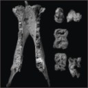
Additions to the elasmobranch assemblage from the Bandah Formation (middle Eocene, Bartonian), Jaisalmer District, Rajasthan, India, and the palaeobiogeographic implications of the fauna
Published online: 23/06/2021
Keywords:
Chondrichthyes; Elasmobranchii; Indian Ocean; Palaeogene; South Asia
https://doi.org/10.18563/pv.44.2.e1
Abstract
Isolated elasmobranch teeth (sharks and rays) from the middle Eocene (Bartonian) Bandah Formation in the Jaisalmer District of Rajasthan, India are described. The remains improve our knowledge of the environment represented by this lithostratigraphic unit and the ecology preserved therein. Seventeen unequivocal taxa were identified, including Nebrius sp., Striatolamia aff. S. macrota, Brachycarcharias atlasi, B. lerichei, cf. Jaekelotodus sp., Carcharhinus mancinae, Rhizoprionodon sp., Physogaleus sp., Galeocerdo clarkensis, G. eaglesomei, Odontorhytis aff. O. pappenheimi, “Rhinobatos” sp., “Dasyatis” sp., Coupatezia sp., “Aetomylaeus” sp., “Rhinoptera” sp., and Ouledia aff. O. lacuna. Of these, “Aetomylaeus” sp., B. atlasi, C. mancinae, G. clarkensis, G. eaglesomei, cf. Jaekelotodus sp., Nebrius sp., Odontorhytis aff. O. pappenheimi, Ouledia aff. O. lacuna, and “Rhinoptera” sp. are reported from the middle Eocene of India for the first time. The Bandah Formation elasmobranch palaeofauna has close affinities to the Palaeocene-Eocene Tethyan/Paratethyan faunas of Africa, Madagascar, Asia, and Europe, and some taxa indicate a western hemisphere influence from North America. The Bandah Formation palaeofauna indicates that deposition occurred in a moderately shallow marine environment. The Bartonian age is primarily based on foraminifera but is corroborated by the presence of elasmobranch taxa that also occur in contemporaneous deposits elsewhere. The marine regression started during the early Palaeogene, and our study indicates that the sea completely withdrew from the Jaisalmer Basin after the deposition of the Bandah Formation. This event may have been synchronous with the middle Eocene uplift of the Himalayan-Tibetan Plateau.
PV article infos
Published in 44-2 (2021)
|
PDF
|
|
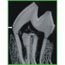
Enamel hypoplasia on rhinocerotoid teeth: Does CT-scan imaging detect the defects better than the naked eye?
Published online: 03/01/2022
Keywords:
fossil teeth; method; micro-CT imaging; Rhinocerotoidea
https://doi.org/10.18563/pv.45.1.e2
Abstract
Micro-CT imaging is an increasingly popular method in paleontology giving access to internal structures with a high resolution and without destroying precious specimens. However, its potential for the study of hypoplasia defects has only recently been investigated. Here, we propose a preliminary study to test whether hypoplastic defects can be detected with micro-CT (μCT) scan and we assess the costs and benefits of using this method instead of naked eye. To do so, we studied 13 fossil rhinocerotid teeth bearing hypoplasia from Béon 1 (late early Miocene, Southwestern France) as positive control and 11 teeth of the amynodontid Cadurcotherium (Oligocene, Phosphorites du Quercy, Southwestern France), for which enamel was partly or totally obscured by cement. We showed that all macroscopically-spotted defects were retrieved on 3D reconstructions and selected virtual slices. We also detected additional defects using μCT scan compared to naked eye identification. The number of defects detected using μCT was greater in the Cadurcotherium dataset (paired-sample Wilcoxon test, p-value = 0.02724) but not for our control sample (paired-sample Wilcoxon test, p-value = 0.1171). Moreover, it allowed for measuring width and depth of the defects on virtual slices (sometimes linked to stress duration and severity, respectively), which we could not do macroscopically. As μCT imaging is both expensive and time consuming while not drastically improving the results, we recommend a moderate and thoughtful use of this method for hypoplasia investigations, restricted for instance to teeth for which enamel surface is obscured (presence of cement, uncomplete preparation, or unerupted germs).
PV article infos
Published in 45-1 (2022)
|
PDF
S.I. Data
|
|
Dortokid turtle remains from the Upper Cretaceous of Cruzy (Hérault, southern France) and phylogenetic implications
Published online: 14/11/2022
Keywords:
Cruzy; Dortoka vasconica; France; Late Cretaceous; Turtle
https://doi.org/10.18563/pv.45.2.e3
Abstract
An isolated right costal 1 from the Late Cretaceous Massecaps locality (Cruzy, Hérault, southern France) is assigned to Dortoka vasconica (Dortokidae). This find adds a new element to the Late Cretaceous turtle fauna of Cruzy and further supports the hypothesis that two distinct lineages of Dortokidae were present in Europe during the Late Cretaceous-Paleogene due to geographical isolation.
PV article infos
Published in 45-2 (2022)
|
PDF
|
|
First report of Cylindracanthus (Osteichthyes) from the Eocene of India
Published online: 25/03/2024
Keywords:
Cylindracanthus; Eocene; histology; rostrum; Umarsar mine.
https://doi.org/10.18563/pv.47.1.e2
Abstract
Fossils of the endangered sturgeons and peddlefishes are widely distributed. We here report for the first time the presence of one of the extinct osteichthyes genus Cylindracanthus (Liedy 1856a) from the Early Eocene lignite-bearing successions of the Kutch Basin, India. The present well preserved rostrum is characterised by numerous wedge-shaped components encircling the central canal that runs along its length, paired at the base and each wedge contributing to the formation of a ridge. The rostrum lacks teeth. The present find extends the palaeobiogeographical distribution of Cylindracanthus considerably and supports its Eocene age as dental remnants preserved in Cylindracanthus sp. shows a decrease in remanent dentition and tooth bases from the Cretaceous to the Eocene. Cylindracanthus is an useful palaeoenvironmental indicator as it has been found associated typically with deposits of nearshore marine environments.
PV article infos
Published in 47-1 (2024)
|
PDF
|
