
|
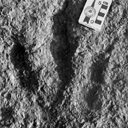
Les traces de pas de Dinosaures et autres Archosaures du Lias inférieur des grands Causses, Sud de la France
Published online: 15/12/2002
Keywords:
Dinosauroid footprints; France; Grands-Causses; Hettangian; ichnostratigraphy; paleoenvironments; Sinemurian; statistical results
https://doi.org/10.18563/pv.31.1-4.1-143
Abstract
The Causses" is a near 3400 km2 large plateau located in the south of France. Here the first dinosaur footprints where found in 1935. After this, this area has yielded an ever-increasing number of ichnites now in excess of 500 specimens. These latter, 15 to 50 cm long, tridactyl or tetradactyl footprints of generally biped animals, were discovered at the surface of Hettangian to lower Sinemurian dolomite layers within 4 distinct stratigraphic units. The 35 sites bearing ichnites are located on the plateau margin. For the first time, morphologic characters studied through descriptive statistic methods with the usual parameters and classical Student and Snédecor tests, allowed us, to divide the whole set of biped traces into 6 ichnospecies. Their definitions are further constrained by multivariate statistical results using Principal Component Analysis (PCA), Factor Analysis of correspondances (FAC) and Discriminant Analysis (DA). All have confirmed the morphologic observations. So that now, the following taxa are identified : Grallator variabilis, G. lescurei, G. sauclierensis, G. minusculus, Eubrontes giganteus, Dilophosauripus williamsi, cf. Moraesichnium, Orníthopus fabrei nov ichnosp. The more immediately visible differences relate to the interdigital II-IV divarication and the digit length ratio. To this panel, we must add Batrachopus deweyi and shapes suggesting Trisauropodichnus and/or Anomoepus. Among all ichnite associations described in the lower Liasic, the New England assemblage presents the most affinities with ours. It shows the ichnotaxa Grallator, Dilophosauripus, Eubrontes, Batrachopus without forgetting Ornithopus fabrei nov. ichnosp. which is close to Ornithopus gallinaceus from the Massachusetts and Connecticut basins. On comparing the present early Jurassic ichnofauna of the Causses with the ones of the Middle and Upper Triassic formations of the eastem border of the Massif Central (France), it appears that tridactyl footprints become more and more numerous and large from Triassic to Early Jurassic. In the Causses, these latest are prevalent but in Quercy (France), Poland, Italy, USA, they are also associated with Omithopoda, Thyreophora and Sauropoda ichnites. Footprint areas considered here were generaly under an arid climate. Animals that passed by were heavy and bulky possible Megalosaur trackmakers, and lighter and slender Coelophysids or Ceratosaurs. For all, these areas were pathways as the orientations of the trackways seem point out. The directions followed by these reptiles were without any important variation during the Hettango-Sinemurian stages. These areas were also used from time to time by Crocodilomorpha and may be tetradactyl (I-IV) bipedal avian Theropods. However, the number of such trackways in sites, sometimes substantial, should not lead us to overestimate the trackmakers populations. These last were probably relatively moderately abondant in this inter-supratidal swamp environment. In the Causses, ichnites are connected with former algo-laminated deposits (Algal mats) which were rapidly hardened by means of calcitisation of cyanobacteria. The result has been a moderate depth of footprints; autopodia disturbing only a few cm of the carbonate substrate. Other fossils have been discovered : invertebrates with thin bivalve and gastropod shells, crustaceans tests and plants. These latter suggest the existence of paleomangroves like environments but also continental vegetation periodically overruning the swamp environment during regression/transgression cycles. At these times, wooded parts of it, could become protecting, feeding, resting and nesting places.
PV article infos
Published in Vol. 31, Fasc. 1-4 (2002)
|
PDF
|

|
The Gliridae (Mammalia) from the oligocene (MP24) of Gröben 3 in the folded molasse of southern Germany
Published online: 28/12/2001
Keywords:
Biostratigraphy; Cyrena Beds; folded molasse; Germany; Gliridae; level MP 24; Mammals; Oligocene; Palaeoecology
Abstract
This study describes four taxa of Gliridae from the Oligocene mammal locality Gröben 3: Gliravus tenuis BAI-ILO, 1975, Bransatoglis micio (MISONNE, 1957), B. planus (BAHLO, 1975) and B. heissigi n. sp. Gliravus tenuis from Gröben 3 is somewhat more advanced than the type population found in Heimersheim. This confirms previous research suggesting that Gröben 3 should be dated earlier than Heimersheim (MP 24). The first documented occurrence of B. mício around level MP 24 was found in Gröben 3. An abundance of tooth material from B. planus in Gröben 3 makes it possible, for the first time, to observe evolutionary stages within this species from MP 21 until MP 28. B. heissigi n. sp. is restricted to level MP 24. This species is located between B. mísonnei (MP 20 - 23) and Microdyromys praemurinus (MP 25 - 28). Within the lineage Bransatoglis bahloi - B. misonnei - B. heissigi, a decrease in size is noticeable.
PV article infos
Published in Vol. 30, Fasc. 3-4 (2001)
|
PDF
|
|
Nouvelles données sur les Ichnites de dinosaures d'El Bayadh (Crétacé Inférieur, Algérie)
Published online: 16/12/2008
Keywords:
Algeria; Brezina; El Bayadh; Ichnites; Lower Cretaceous; Sauropoids; Theropoids
https://doi.org/10.18563/pv.36.1-4.7-35
Abstract
Evidence of 350 Lower Cretaceous Dinosaur footprints is pointed out in El Bayadh area. Their preliminary study allow to distinguish four trackway assemblages which reveal vertebrate bipedal presence forms of tri-and tetradactylous Dinosauroïds (Assemblages 1-3) and quadrupidal Sauropoïd (Assemblage 4).
The analysis of their footprint biometric features will attribute the quadrupidal Sauropoïd form to Brontopodus ichnogenus which is weIl known in the Jurassic and Cretaceous periods. In retum and despite their age, the dinosauroïd forms were approached, temporarily, to Grallator and Eubrontes types.
The occurrence of the dinosaur traces (Theropoïd and Sauropoïd) constitutes, in the Lower Cretaceous, an important first step of the knowlege of the marshy Reptilian fauna which takes over, from the begining of the Secondary Era, a wide paleogeographie area on the Southem Tethyan margin.
PV article infos
Published in Vol. 36, Fasc. 1-4 (2008)
|
PDF
|
|
A reassessment of the giant birds Liornis floweri Ameghino, 1895 and Callornis giganteus Ameghino, 1895, from the Santacrucian (late Early Miocene) of Argentina.
Published online: 13/12/2016
Keywords:
Argentina; Aves; Callornis; Liornis; Miocene
https://doi.org/10.18563/pv.40.2.e3
Abstract
The status of the giant bird taxa Liornis floweri and Callornis giganteus from the Santa Cruz Formation (late Early Miocene) of Patagonia, first described by Ameghino (1895) is reassessed on the basis of a re-examination of the type material at the Natural History Museum, London. Liornis floweri, which lacks a Pons supratendineus on the tibiotarsus and has an unbifurcated Canalis interosseus distalis on the tarsometatarsus, is clearly a brontornithid and is considered as a junior synonym of Brontornis burmeisteri. Ameghino’s replacement of Callornis by Eucallornis is unjustified. Callornis giganteus is a chimera based on a phorusrhacid tarsometatarsus (probably belonging to Phorusrhacos longissimus) and a brontornithid tibiotarsus. The latter can be considered as the lectotype of Callornis giganteus, which may represent a small morph of Brontornis burmeisteri or a distinct taxon. It is referred to here as Brontornithidae indet. The tarsometatarsus described by Dolgopol de Saez (1927a,b) as Liornis minor and considered by her as a gracile brontornithid apparently has a bifurcated Canalis interosseus distalis and should therefore be placed among the Phorusrhacidae.
PV article infos
Published in Vol.40-2 (2016)
|
PDF
|

|
A new rodent from Quaternary deposits of the Canary Islands and its relationships with Neogène and recent murids of Europe and Africa.
Published online: 15/12/1988
Keywords:
Canary Islands; Holocene; Island evolution; Muridae; PHYLOGENY; Rodents; Spain
https://doi.org/10.18563/pv.18.4.241-262
Abstract
A peculiar new rodent, Malpaisomys insularis nov. gen., nov. sp., is described from subfossil deposits of the eastern Canary Islands. The species shows some highly specialized skull features although its molars exhibit a mixture of primitive and derived characters among which a partial stephanodonty is most notable. A comparison of the new rodent with several Miocene to Holocene Muridae shows that Malpaisomys possibly shares a common ancestor with Acomys and Uranomys.
PV article infos
Published in Vol. 18, Fasc. 4 (1988)
|
PDF
|

|
Pantolestidae nouveaux (Mammalia, Insectivora) de l'Eocène moyen de Bouxwiller (Alsace).
Published online: 31/03/1970
Keywords:
Bouxwiller; Insectivora; Mammalia; Middle Eocene; Pantolestidae
https://doi.org/10.18563/pv.3.3.63-82
Abstract
The Pantolestidae from the middle eocene of Bouxwiller are the subject of a detailed study. Buxolestes hammeli (n. g., n. sp.) is not closely related to any other European or North American form described until now; it presents, however, some characters in common with Pantolestes, a form of the same age from North America. A parallel evolution from a common ancestral form could explain this ressemblance.
Another form (gen. and sp. indet.) accompanies Buxolertes hammeli in the Bouxwiller fauna.
PV article infos
Published in Vol. 03, Fasc. 3 (1970)
|
PDF
|

|
Avant-propos
Published online: 16/12/1996
Keywords:
D.E.Russell
Abstract
Le présent volume est l'aboutissement d'un projet né il y a presque cinq ans. En décembre 1991, l'un d'entre nous (MG) prenait des contacts en vue de proposer un symposium sur les mammifères fossiles, dédié à D.E. Russell, dans le programme du 4e Congrès de la European Society for Evolutionary Biology. Ce congrès, baptisé "Evolution 93", devait se tenir à Montpellier en août 1993. Son Comité d'Organisation, animé par F. Catzeflis, recherchait des organisateurs de symposiums. L'idée fut acceptée avec enthousiasme par le second d'entre nous (PDG), et le titre de notre Symposium fut précisé: " Palaeobiology and Evolution of Early Cenozoic Mammals - A Symposium in Honor of D.E. Russell". Le projet fut formellement accepté par le Comité d'Organisation en avril 1992.
PV article infos
Published in Vol. 25, Fasc. 2-4 (1996)
|
PDF
|

|
Contribution à la classification des Pistes de Vertébrés du Trias : les types du Stormberg d'Afrique du Sud (2 ème Partie: le Stormberg supérieur - 1. Le biome de la zone B/1 ou niveau de Moyeni: ses biocénoses).
Published online: 01/12/1974
Keywords:
biocenosis; Footprints; South Africa; Stormberg; Trias
https://doi.org/10.18563/pv.6.ext
Abstract
Les Pistes de Vertébrés du Stormberg Supérieur ("Trias terminal à Rhétien"), ou Quthingien
Si les zones du Stormberg inférieur se sont révélées contenir de nombreuses traces, surtout dans les faciès dits "Molteno moyen et supérieur", représentant apparemment la base du Keuper, il est frappant de voir pratiquement l'ensemble de cette grosse faune "Molteno" disparaître avec la fin de cette période, que nous avons appelée le "Maphutsengien".
Dès les premières zones du Stormberg supérieur, que nous nommons le"Quthingien" la zoocénose et la phytocénose, en même temps que les données d'ensemble manifestées par l'environnement, sont modifiées. Nous ne verrons plus guère de dépôts marécageux à flore riche et variée, parfois même luxuriante. Les fougères elles-mêmes ont disparu. Elles sont remplacées par de maigres plantes, aux feuilles très souvent filiformes qui paraissent témoigner d'un climat continental. Le sol est devenu de plus en plus rouge, avec des variations latérales beaucoup plus accusées. Les fleuves amenant des galets des monts du "Grand Sud" ont tari. La faune va en subir les conséquences. Certaines des espèces se révèleront sautillantes ou coureuses, pour un grand nombre plus légères et pour la quasi-totalité d'apparence carnivore ou entomophages, les phytophages devant se contenter d'un régime ingrat,difficile ou à tout le moins irrégulier,les dépôts le montrent.
C'est dans ces conditions que s'inaugure notre Etage nouveau,quelque peu discordant sur les zones A/5, A/6 ou A/7 du Stormberg inférieur (Maphutsengien). Le Stormberg supérieur (ou Quthingien) commence avec le paléopaysage remarquable dit de Moyeni, que nous allons maintenant étudier, typologiquement, avec ses homologues du même âge. Quelques 38 types d'animaux tous nouveaux vont défiler à nos yeux lors de la zone de base de cet Etage, ou zone B/1.
L'on nous avait proposé d'intituler ce Ile Tome de la série : "La grande Dalle de Moyeni et ses homologues. Paléo-spectacles, scènes et paysages animaux au Lesotho à l'approche du Trias finissant". Nous avons préféré garder le sous-titre plus haut, peut-être plus prosaïque.
Un llle Tome est en préparation : "Les développements ultérieurs et terminaux de la faune du Gondwana".
PV article infos
Published in Vol. 6, Ext (1974)
|
PDF
|

|
Nouveaux Mammifères Eocènes du Sahara Occidental
Published online: 01/11/1979
Keywords:
Eocene; Mammals; Occidental Sahara
https://doi.org/10.18563/pv.9.3.83-115
Abstract
The fossil mammals collected from the Eocene of Hammada du Dra (northwest Sahara. Algeria) and two fragmentary teeth from the Lutetian of M'Bodione Dadere (Senegal) are described.
The fossils from the northwest Sahara come from a lacustrian deposit dated by charophytes (Raskyella aff. pecki, Raskyella n. sp.. Maedleriella lavocati, Maedleriella sp. et ? Peckichara sp.) as Middle Eocene or perhaps Lower Eocene (Gevin, Feist and Mongereau, 1974). Several hyracoids (3 or 4) identified from this formation extends the age of the family Pliohyracidae Osborn in Africa. Three forms appear to belong in the genera Megalohyrax, Titanohyrax and perhaps Bunohyrax which have been know until now only from the lower Oligocene of the Fayum (M. gevini n. sp. ; T. mongereaui n. sp.. ? Bunohyrax or Megalohyrax indet.). Another hyracoidof small size is referred to a new genus, Microhyrax (M. lavocati n. sp.).
Helioseus insolitus n. g. n. sp. is described without ordinal assignment. Azibius (Sudre, 1975) which has been the subject of questions and interpretations is reviewed.
Only one tooth from the Lutetian of M'Bodione Dadere is complete enough to interpret. lt probably belongs to a condylarth and demonstrates for the first time, the presence of the order in Africa. The second tooth is too fragmentary for comment.
In conclusion., the paleobiogeographic role of Africa at the end of the cretaceous and the beginning of the Cenozoic is discussed.
PV article infos
Published in Vol. 09, Fasc. 3 (1979)
|
PDF
|

|
Les mammifères Montiens de Hainin (Paléocène moyen de Belgique) Part1: Multituberculés.
Published online: 01/11/1979
Keywords:
Belgium; Hainin; Mammals; multituberculates; Paleocene
https://doi.org/10.18563/pv.9.4.117-131
Abstract
The Montian locality of Hainin (Hainaut, Belgium) yielded about twenty teeth of Multituberculates. They are very peculiar forms, showing no affinities, at the generic level, with those hitherto known from North America, Asia and Europe. They are referred to the new taxa Boffius splendidus nov. gen., nov. sp., Hainina belgica nov. gen., nov. sp., and H. godfriauxi nov. gen., nov. sp. They expose some common features, such as the advanced type of first upper molar. possessing at least three complete rows of cusps. Because of this, and also of the upper premolar reduction, Boffius splendidus appears as the most specialized form within the Ptilodontoidea suborder.
Several other characters of Hainina seem to be less advanced, such as the great number of upper premolars and the simple cusp-formula of the first lower molar.
Till now, only H. godfriauxi has been recovered within the Thanetian fauna from Cemay-les-Reims, where it is very poorly documented.
PV article infos
Published in Vol. 09, Fasc. 4 (1979)
|
PDF
|

|
Die Ohr-Region der Paulchoffatiidae (Multituberculata, Ober-Jura).
Published online: 15/11/1988
Keywords:
Multituberculata; Ober-Jura; Paulchoffatiidae; Petrosum; Portugal
https://doi.org/10.18563/pv.18.3.155-185
Abstract
The petrosal of the Paulchoffatiidae HAHN, 1969 is described and compared with that of younger multituberculates and of other Mesozoic mammals. The "Morrison petrosal", described by Prothero (1983), is also discussed; it probably belongs to the multituberculates. The reconstruction of the ventral side of the Paulchoffatiinae-skull, given by Hahn in 1987, is completed by addition of the otic and the occipital region.
PV article infos
Published in Vol. 18, Fasc. 3 (1988)
|
PDF
|

|
Les rongeurs de l' Eocène inférieur et moyen d'Europe Occidentale; Systématique, phylogénie, biochronologie et paléobiogéographie des niveaux-repères MP 7 à MP 14.
Published online: 15/12/1999
Keywords:
Biochronology; Early and Middle Eocene; Gliridae; Ischyromyidae; Mammalia; MP Scale; New Genus and Species; Palaeogeography; PHYLOGENY; Rodents; Theridomyidae; Western Europe
Abstract
Fourteen distinct phyletical lineages which belong at least in three families: Ischyromyidae ALSTON, 1876, Gliridae THOMAS, 1896 and Theridomyidae ALSTON, 1876, have been identified after the study of more than 3600 rodent dental remains from about twenty Early and Middle Eocene european localities. A systematical and phylogenetical revision of these rodents has been achieved. Nearly all the specific and generic diagnosis are emended. Several new combinations and synonymies are proposed. Four new species and two new genera, Euromys nov. (Ailuravinae) and Hartenbergeromys nov. (Microparamyini), are named and described. Euromys nov. gen. is known by three distinctive ypresian (MP 7 to MP 10 european reference levels) chronospecies. This new lineage is thought to be the direct ancestor of Meldimys MICHAUX, 1968 and Ailuravus RUTIMEYER, 1891. A new species of the genus Plesiarctomys BRAVARD, 1850, Pl. lapicidinarum from Condé-en-Brie (MP 8-9 reference level), allows to relate the Plesiarctomys lineage to the Pseudoparamys MICHAUX, 1964 one. The taxa Sparnacomys HARTENBERGER, 1971, Pantrogna HARTENBERGER, 1971, and Corbarimys MARANDAT, 1989 are erected to genus rank; the last one is not thought to be an Ischyromyidae. A new chronospecies of Pantrogna, P. marandati nov. sp. from the locality of Prémontré (MP 10 reference level), is described. This lineage is at the origin of two others, namely Masillamys TOBIEN, 1954, including M. mattaueri (HARTENBERGER, 1975) nov. comb. (MP 10 reference level), and Hartenbergeromys nov. gen., known from MP 10 (H. hautefeuillei nov. sp.) and MP 11 (H. parvus TOBIEN, 1954) reference levels. The phylogenetical position of Hartenbergeromys nov. gen., at the origin of the european family Theridomyidae, is discussed. The systematical and phylogenetical status of two probable Paramyinae, "Paramys" woodi MICHAUX, 1964 and an unnamed genus and species, are discussed. New populations of the primitive Gliridae Eogliravus HARTENBERGER, 1971 and of the primitive Theridomyidae Protadelomys HARTENBERGER, 1968, are described and assigned to previously known species.
PV article infos
Published in Vol. 28, Fasc. 2-4 (1999)
|
PDF
|

|
Compléments sur les Chiroptères de l'Eocène moyen d'Europe. Les genres Palaeochiropteryx et Cecilionycteris.
Published online: 01/10/1980
Keywords:
Chiroptera; Geiseltal; Messel; Middle Eocene
https://doi.org/10.18563/pv.9.ext.91-126
Abstract
New dental and skeletal material referable to Palaeochiropteryx tupaiodon from the Middle Eocene locality of
Messel (G.F.R.) is studied, which provides additions to the previously gained knowledge of this european genus. Dental specimens from Geiseltal (G.D.R.), also of Middle Eocene age, allow us to analyze Cecilionycteria prisca. Some of these are separated to establish a new genus, Matthesia, and two new species, M. germanica and M. ? insolita.
PV article infos
Published in Vol. 9, Ext (1980)
|
PDF
|

|
Données nouvelles sur le genre Stehlinia (Vespertilionoidea, Chiroptera) du Paléocène d'Europe
Published online: 01/12/1974
Keywords:
Chiroptera; Palaeocene; Vespertilionoidea
https://doi.org/10.18563/pv.6.3-4.253-272
Abstract
Cet article présente une étude détaillée du genre Stehlinia, un chiroptère du Paléogène d'Europe, basé sur un matériel abondant issu notamment du gisement d'Escamps (Quercy). L'analyse révèle que Stehlinia possède un mélange de caractères primitifs (comme une denture tribosphénique complète et des prémaxillaires soudés) et évolués (notamment dans le squelette post-crânien), le rapprochant des vespertilionoïdes actuels, en particulier des Kerivoula.
PV article infos
Published in Vol. 06, Fasc. 3-4 (1975)
|
PDF
|

|
Nouvelles données sur les mammifères du Thanétien et de l'Yprésien du bassin d'Ouarzazate (Maroc) et leur contexte stratigraphique.
Published online: 15/12/1998
Keywords:
early Paleogene; magnetostratigraphy; Mammals; Morocco; North Africa; Ouarzazatz basin; Systematics
Abstract
New faunal and stratigraphical data on the vertebrates localities from the early Paleogene of the Ouarzazate Basin (Adrar Mgorn 1, Adrar Mgorn 1 bis et N'Tagourt 2), Morocco, are presented. A magnetostratigraphical study, the first for such early Paleogene Arabo-African mammal localities, and the discovery of probable remains of the nannofossil Discoaster support the Thanetian age of the Adrar Mgorn 1 site. The magnetostratigraphy suggests a slightly later age than was thought for the Paleogene formations of the local series of Tinerhir and for the vertebrate localities: late or latest Thanetian for Adrar Mgorn 1 and Adrar Mgorn 1 bis, middle Ypresian for N'Tagourt 2. It also indicates a lower position of the KT boundary in the series. Two tons of matrix recovered in the vertebrate sites have vielded new data on the micromammals. A damaged lower molar from N'Tagourt 2 is referable to Khamsaconus bulbosus and supports the proboscidean affinities of this species and especially possible relationships with bunolophodont taxa such as elephantiforms. A lower molar from Adrar Mgorn 1 bis belongs to a new form which can be identified as a plesiadapiform or an euprimate close to Altiatlasius koulchii though significantly larger. A new material from Adrar Mgorn 1 illustrates a new dilambdodont adapisoriculid species which is referable to Garatherium : ?Garatherium todrae n. sp. Another species referred to Garatherium is known in the locality (?Garatherium n. sp.). Garatherium is a new lineage from the Ouarzazate basin which crosses the Paleocene-Eocene boundary together with Palaeoryctes, Didelphodontinae gen. and sp. 2, Todralestes, and Afrodon, and it is the first Paleocene-Eocene lineage identified outside of this basin (Garatheríum is based on a species from El Kohol, Algeria). Among the Paleocene-Eocene lineages from the Ouarzazate basin, it should be also mentioned a new possible carnassial form (carnivoran or creodont; Adrar Mgorn 1), and an upper molar of Cimolestes cf. incisus (Adrar Mgorn 1 bis). The upper molar THR 168 previously reported as from an indeterminate didelphodontine is here identified as the M1/ of Afrodon chleuhi. The micromammal faunas from the Ouarzazate basin are positioned in the global chronological framework of the mammal localities from the Paleogene of the Arabo-African domain.
PV article infos
Published in Vol. 27, Fasc. 3-4 (1998)
|
PDF
|

|
Contributions à l'étude du gisement Miocène supérieur de Montredon (Hérault). Les grands mammifères. 7 - Les proboscidiens Deinotheriidae
Published online: 15/11/1988
Keywords:
allometry; Astaracian; Deinotherium; Montredon; Systematics; taphonomy; Vallesian
https://doi.org/10.18563/pv.18.ext.135-175
Abstract
Some complete tooth rows and about one hundred isolated teeth enabled the identification of the deinothere of the Vallesian site Montredon (Hérault) as Deinotherium giganteum KAUP 1829, mainly by comparisons with the likewise Vallesian sample of the type locality Eppelsheim (Rheinhessen, F.R.G.).
Scatterdiagrams of the teeth show the importance of allometry during the phyletic size increase of the European deinotheres.
Some taphonomic problems of the Montredon deinothere are briefly mentioned.
PV article infos
Published in Vol. 18, Ext (1988)
|
PDF
|

|
La poche à phosphate de Ste-Néboule (Lot) et sa faune de vertebres du Ludien supérieur. 7- Didelphides (Marsupiaux)
Published online: 25/09/1978
Keywords:
Eocene; Quercy Phosphorites
https://doi.org/10.18563/pv.8.2-4.231-242
Abstract
The family Didelphidae is represented by three species in the Sainte-Néboule site, phosphorites of Quercy (lower Oligocene, San Cugat's nivel): Amphiperatherium minutum (Aymard), Amphiperatherium sp. and Peratherium cuvieri (Fischer). Only the first and third species are abundant (88 and 97 pieces). This two populations are described. The marsupial fauna of the european lower Oligocene is not recognized in its entirety in this site.
PV article infos
Published in Vol. 08, Fasc. 2-4 (1978)
|
PDF
|
|
A new Desmodillus (Gerbillinae, Rodentia) species from the early Pliocene site of Langebaanweg (South-western Cape, South Africa)
Published online: 20/01/2017
Keywords:
Lower Pliocene; Muridae; Rodentia; RSA
https://doi.org/10.18563/pv.41.1.e1
Abstract
Situated in the Cape region of the Republic of South Africa (RSA), the paleontological site of Langebaanweg is dated to 5.1 Myr and is famous for having yielded an abundant vertebrate assemblage, including numerous rodent species from the Mio-Pliocene transition. Based on molar morphology and skull anatomy, the single Gerbillinae taxon identified at Langebaanweg and described in this paper is allocated to Desmodillus, which is a modern monotypic South African endemic genus. It is significant in being the oldest representative of the genus in Africa. We describe here a new species of this genus which is larger than the modern D. auricularis, but nevertheless retains some of its main characteristics, namely the shape of the maxilla and mandible, the presence of poorly fused alternating cusps, and no longitudinal crest. This taxon differs from modern South African Gerbilliscus representatives in some mandibular and maxillary characters, in the m1 prelobe cusp, and in having less fused cusps. Two fossil Gerbillinae discovered in the Upper Miocene of Africa and Asia, Abudhabia and Protatera, have been compared with the new species. We discuss their relationships with modern and Plio-pleistocene Gerbillinae and conclude that Abudhabia could be the sister taxon of Desmodillus and that around 6-5 Myr a vicariance event allowed Gerbillinae to diversify into modern Desmodillus in South Africa, and Gerbilliscus in East Africa. The murine/gerbilline ratio, which is a good indicator of rainfall, supports other proxies which suggest that at 5.1 Myr the climate in the Langebaanweg region was more humid than today.
PV article infos
Published in Vol 41-1 (2018)
|
PDF
S.I. Data
|

|
An Australian Miocene Brachipposideros (Mammalia, Chiroptera) related to Miocene representatives from France
Published online: 15/12/1982
Keywords:
Australia; bats; Chiroptera; Miocene
https://doi.org/10.18563/pv.12.5.149-172
Abstract
A new middle Miocene hipposiderid bat is described from a limestone deposit on Riversleigh Station in north-western Queensland. Hipposideros (Brachipposideros) nooraleebus n. sp. is the first record of this subgenus from anywhere in the world outside of France. The palaeoecological setting of the fossil bats appears to have been a relatively quiet, sunny lime-enriched tropical pool that contained tortoises, crocodiles and fish. It is possible that the bats were washed into the pool from an adjacent cave.
The Riversleigh bat most closely resembles the French Burdigalian (early middle Miocene) bat H. (B.) aguilari. It is also possible that it may have been closely related to the original Australian hipposiderid stock that ultimately gave rise to the endemic monotÿpic Rhinonycteris aurantius. The disjunct distribution of species of H. (Brachipposideros) suggests that representatives of this subgenus will be found in at least tropical southern Asia.
PV article infos
Published in Vol. 12, Fasc. 5 (1982)
|
PDF
|

|
Contributions à l'étude des micromammifères du gisement Miocène supérieur de Montredon (Hérault). 4 - Les chiroptères
Published online: 30/06/1982
Keywords:
Chiroptera; Hérault; Late Miocene; Micromammals; Montredon
https://doi.org/10.18563/pv.12.3.133-140
Abstract
The Montredon local fauna yielded very rare bats, represented by damaged isolated teeth. Only a few documents are available for this period of the European Neogene. ln this poor state of knowledge, the material represents three undetermined species, a supposed molossid and two vespertilionids.
PV article infos
Published in Vol. 12, Fasc. 3 (1982)
|
PDF
|
