
Pterosaurs from Coahuila
Abstract book of the 18th Conference of the EAVP
Two enigmatic rodents from Lavergne (MP 16), Quercy Phosphorites
Les sélaciens du Miocène de la région de Montpellier
Muridae du Pliocène supérieur d'Espagne et du midi de la France.
Les Chiroptères du Miocène inférieur de Bouzigues. 1- Etude systématique.
Eocene (57) , Quercy Phosphorites (38) , Systematics (32) , Rodents (29) , Mammalia (27) , Rodentia (25) , Miocene (24)

|
Rythme et modalités de l'évolution chez les rongeurs à la fin de l'Oligocène-leurs relations avec les changements de l'environnement.Bernard ComtePublished online: 15/12/2000Keywords: Environment; evolution; Oligocene; Rodents; Systematics Abstract The analysis of oxygene isotope variations as well as paleobotanical data suggest that the Oligocene/Miocene boundary corresponds to a transitional period marked by floristical and climatic variations. During this period, the pyreneo-alpine tectonics has contribued to modify the geography and western Europe landscapes. Faunal changes (appearances, extinctions, migrations) are observed in different mammalian groups, notably in the rodents. A study of the evolutionary trends and patterns in paleogene rodents is involved for the period ranging from level MP 28 of the Late Oligocene to the Early Miocene, including the Oligo-Miocene boundary. PV article infos Published in Vol. 29, Fasc. 2-4 (2000) |
|

|
Nouvelle quantification de l'Hypsodontie chez les Theridomyidae : l'exemple de Theridomys ludensis nov. sp.Monique Vianey-Liaud
Published online: 30/12/1985 |
|

|
Fossil snakes from the Palaeocene of Sao José de Itaborai, Brazil.Part 1 Madtsoiidae, Aniliidae.Jean-Claude Rage
Published online: 15/12/1998 |
|

|
Eggshell microstructure and porosity of the Nicobar scrubfowl (Megapodius Nicobariensis, great Nicobar island, India)Géraldine Garcia
Published online: 16/12/2008 |
|
|
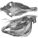
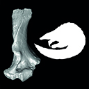
Old world hemiones and new world slender species (Mammalia, Equidae)Véra Eisenmann, John Howe and Mario PichardoPublished online: 16/12/2008Keywords: Amerhippus; biometry; Equus; Holocene; New World; Old World; Osteology; Pleistocene; Pliocene https://doi.org/10.18563/pv.36.1-4.159-233 Abstract Morphological and biometrical description of skulls, teeth, and limb bones of extant and fossil Old World herniones (including E. hydruntinus) and of New World 'stilt-Iegged' and other slender species from Blancan to Holocene. An Appendix presents ways in which the approximate size of some missing bones or dimensions may be deduced from available ones. PV article infos Published in Vol. 36, Fasc. 1-4 (2008) |
|

|
Les Dipodidae (Mammalia, Rodentia) d'Europe occidentale au Paléogène et au Néogène inférieur: origine et évolution.Marguerite Hugueney and Monique Vianey-Liaud
Published online: 01/10/1980 |
|

|
La poche à phosphate de Ste-Néboule (Lot) et sa faune de vertébres du Ludien Supérieur. 2- Amphibiens. Etude PreliminaireJean-Claude Rage
Published online: 25/09/1978 |
|

|
Sur la présence de dents de mammifères (Creodonta, Perissodactyla) près de la limite Paléocène-Eocène à Hoegaarden, BelgiqueRichard Smith and Jerry J. HookerPublished online: 16/12/1996Keywords: Belgium; Creodonta; Landenian; Mammals; Perissodactyla Abstract Amongst a collection of selachian teeth made at Hoegaarden in a marine level of Bruxellian (Lutetian) age, containing a reworked Landenian (Sparnacian) fauna mixed with a contemporaneous one, a few teeth of terrestrial mammals have been discovered. They comprise two rare European taxa: ? Hallensia sp. and Palaeonictis gigantea, both known from the Landenian. Even though the ?Hallensia has not been definitely identified, il differs from the only perissodactyl of this age previously recorded from Belgium (Cymbalophus cuniculus). PV article infos Published in Vol. 25, Fasc. 2-4 (1996) |
|

|
Les mammifères Montiens de Hainin (Paléocène moyen de Belgique) Part III : MarsupiauxJean-Yves Crochet and Bernard SigéPublished online: 30/09/1983Keywords: Belgium; Marsupials; Paleobiogeography; Paleocene https://doi.org/10.18563/pv.13.3.51-64 Abstract The oldest european marsupials are described from some specimens (isolated upper molars) recently found from the Hainin sediment (Middle Paleocene of Belgium). These fossils document a new species of the Peradectes genus. They give evidence of a much older occurrence of the marsupials in Europe than it was assumed. They allow us to postulate a didelphid dispersal from South America towards the western-holarctic area operating in two phases : the first one of the Peradectes genus at the end of the Cretaceous; the second one of the Didelphíni tribe at the end of the Paleocene. A central american crossing is likely for the first one, whereas a transafrican way is tentatively argued for the second one. PV article infos Published in Vol. 13, Fasc. 3 (1983) |
|

|
The Gliridae (Mammalia) from the oligocene (MP24) of Gröben 3 in the folded molasse of southern GermanyUndine UhligPublished online: 28/12/2001Keywords: Biostratigraphy; Cyrena Beds; folded molasse; Germany; Gliridae; level MP 24; Mammals; Oligocene; Palaeoecology Abstract This study describes four taxa of Gliridae from the Oligocene mammal locality Gröben 3: Gliravus tenuis BAI-ILO, 1975, Bransatoglis micio (MISONNE, 1957), B. planus (BAHLO, 1975) and B. heissigi n. sp. Gliravus tenuis from Gröben 3 is somewhat more advanced than the type population found in Heimersheim. This confirms previous research suggesting that Gröben 3 should be dated earlier than Heimersheim (MP 24). The first documented occurrence of B. mício around level MP 24 was found in Gröben 3. An abundance of tooth material from B. planus in Gröben 3 makes it possible, for the first time, to observe evolutionary stages within this species from MP 21 until MP 28. B. heissigi n. sp. is restricted to level MP 24. This species is located between B. mísonnei (MP 20 - 23) and Microdyromys praemurinus (MP 25 - 28). Within the lineage Bransatoglis bahloi - B. misonnei - B. heissigi, a decrease in size is noticeable. PV article infos Published in Vol. 30, Fasc. 3-4 (2001) |
|

|
Les traces de pas de Dinosaures et autres Archosaures du Lias inférieur des grands Causses, Sud de la FranceGeorges Demathieu, Georges Gand, Jacques Sciau, Pierre Freytet and Jacques GarricPublished online: 15/12/2002Keywords: Dinosauroid footprints; France; Grands-Causses; Hettangian; ichnostratigraphy; paleoenvironments; Sinemurian; statistical results https://doi.org/10.18563/pv.31.1-4.1-143 Abstract The Causses" is a near 3400 km2 large plateau located in the south of France. Here the first dinosaur footprints where found in 1935. After this, this area has yielded an ever-increasing number of ichnites now in excess of 500 specimens. These latter, 15 to 50 cm long, tridactyl or tetradactyl footprints of generally biped animals, were discovered at the surface of Hettangian to lower Sinemurian dolomite layers within 4 distinct stratigraphic units. The 35 sites bearing ichnites are located on the plateau margin. For the first time, morphologic characters studied through descriptive statistic methods with the usual parameters and classical Student and Snédecor tests, allowed us, to divide the whole set of biped traces into 6 ichnospecies. Their definitions are further constrained by multivariate statistical results using Principal Component Analysis (PCA), Factor Analysis of correspondances (FAC) and Discriminant Analysis (DA). All have confirmed the morphologic observations. So that now, the following taxa are identified : Grallator variabilis, G. lescurei, G. sauclierensis, G. minusculus, Eubrontes giganteus, Dilophosauripus williamsi, cf. Moraesichnium, Orníthopus fabrei nov ichnosp. The more immediately visible differences relate to the interdigital II-IV divarication and the digit length ratio. To this panel, we must add Batrachopus deweyi and shapes suggesting Trisauropodichnus and/or Anomoepus. Among all ichnite associations described in the lower Liasic, the New England assemblage presents the most affinities with ours. It shows the ichnotaxa Grallator, Dilophosauripus, Eubrontes, Batrachopus without forgetting Ornithopus fabrei nov. ichnosp. which is close to Ornithopus gallinaceus from the Massachusetts and Connecticut basins. On comparing the present early Jurassic ichnofauna of the Causses with the ones of the Middle and Upper Triassic formations of the eastem border of the Massif Central (France), it appears that tridactyl footprints become more and more numerous and large from Triassic to Early Jurassic. In the Causses, these latest are prevalent but in Quercy (France), Poland, Italy, USA, they are also associated with Omithopoda, Thyreophora and Sauropoda ichnites. Footprint areas considered here were generaly under an arid climate. Animals that passed by were heavy and bulky possible Megalosaur trackmakers, and lighter and slender Coelophysids or Ceratosaurs. For all, these areas were pathways as the orientations of the trackways seem point out. The directions followed by these reptiles were without any important variation during the Hettango-Sinemurian stages. These areas were also used from time to time by Crocodilomorpha and may be tetradactyl (I-IV) bipedal avian Theropods. However, the number of such trackways in sites, sometimes substantial, should not lead us to overestimate the trackmakers populations. These last were probably relatively moderately abondant in this inter-supratidal swamp environment. In the Causses, ichnites are connected with former algo-laminated deposits (Algal mats) which were rapidly hardened by means of calcitisation of cyanobacteria. The result has been a moderate depth of footprints; autopodia disturbing only a few cm of the carbonate substrate. Other fossils have been discovered : invertebrates with thin bivalve and gastropod shells, crustaceans tests and plants. These latter suggest the existence of paleomangroves like environments but also continental vegetation periodically overruning the swamp environment during regression/transgression cycles. At these times, wooded parts of it, could become protecting, feeding, resting and nesting places. PV article infos Published in Vol. 31, Fasc. 1-4 (2002) |
|

|
Rongeurs nouveaux de l'Oligocène Moyen d'Espagne.Louis ThalerPublished online: 15/09/1969Keywords: Cricetidae; Oligocene; Pseudocricetodon; Rodents; Theridomys https://doi.org/10.18563/pv.2.5.191-207 Abstract Description of four new rodents from a recently discovered locality at Montalban. Theridomys crusafonti nov. sp. is considered as the ancestry of T. Iembronicus. Theridomys varian: nov. sp. includes «Theridomys» morphotypes and «Blainvilllimys» morphotypes; it could be ancestral to B. blainvillei. Pseudoltinomys nanus nov. sp. represents a new lineage paralleling in evolution that of P. gaillardi (which is equally found at Montalban). Pseudocricetodon montalbanensis nov. gen., nov. sp. designates a lineage of very small Cricetidae accompanying Eucricetodon. With these well defined new species and six others present in the locality, Montalban appears as the best faunal reference point within the biochronologic zone of La Sauvetat. PV article infos Published in Vol. 02, Fasc. 5 (1969) |
|

|
Recherches de mammifères paléogènes dans les départements de l'Aisne et de la Marne pendant la deuxième moitié du vingtième sièclePierre LouisPublished online: 16/12/1996Keywords: Biochronology; Eastern Paris Basin; Fossil localities; Mammals; paleoenvironments; Paléogène; Paleogeography Abstract A brief historical account of fossil vertebrate discoveries in the Eastern part of the Paris Basin between the beginning of the nineteenth century and 1950 is given. Other localities discovered since then are presented. A reconstruction of past landscapes is briefly elaborated. A biozonation based on mammals is proposed, from the Late Thanetian to the Middle Bartonian. Paleogeographical considerations are added. Suggestions regarding the search for new marnmal localities are made. PV article infos Published in Vol. 25, Fasc. 2-4 (1996) |
|

|
Mammals and stratigraphy of the continental mammal-bearing Quarternary of South AmericaLarry G. Marshall, Annalisa Berta
Published online: 16/12/1984 |
|

|
Un crane de Chalicothere (Mammalia, Perissodactyla) du Miocène supérieur de Macédoine (Grèce) : remarque sur la phylogénie des ChalicotheiinaeLouis de Bonis
Published online: 14/06/1995 |
|

|
Evolution de la lignée Megacricetodon collongensis-Megacricetotodon roussillonensis (Cricetidae, Rodentia, mammalia) au cours du Midocène inférieur et moyen dans le Sud de la France.Jean-Pierre AguilarPublished online: 14/06/1995Keywords: Cricetids rodents; Evolutionary lineage; Lower and Middle Miocene; Mammalian biochronology; Megacricetodon new species; Southern France Abstract New populations of the genus Megacricetodon have recently been discovered in Southern France.Two new species are defined: M. lemartineli n. sp. and M. fournasi n. sp., their stages of evolution are intermediate between those of M. gersii and M. roussillonensis. Morphological and biometrical analysis indicate the presence of only one lineage: M. collongensis--M. collongensis-gersii--M. gersii--M.lemartineli nov. sp.--M. fournasi nov. sp. and M. roussillonensis. This observation allows to refine the chronology based on rodents, for the Late Early Miocene and the Middle Miocene in the Southern France. PV article infos Published in Vol. 24, Fasc. 1-2 (1995) |
|

|
Les rongeurs du Miocène moyen et supérieur du MaghrebJean-Jacques Jaeger
Published online: 15/05/1977 |
|

|
Les poissons crétacés et tertiaires du bassin des Iullemmeden (République du Niger)Henri Cappetta
Published online: 15/09/1972 |
|

|
Nouveau Dichobunidae (Artiodactyla, Mammalia) du gisement d'Aumelas (Hérault) d'âge Lutétien terminalJean SudrePublished online: 01/10/1980Keywords: Aumelas; Dichobunidae; Hérault; Middle Eocene; Upper Lutetian https://doi.org/10.18563/pv.9.ext.197-211 Abstract The faunal list of the mammals collected at the locality of Aumelas (Hérault, France) is revised. For the first PV article infos Published in Vol. 9, Ext (1980) |
|
|
A pangolin (Manidae, Pholidota, Mammalia) from the French Quercy phosphorites (Pech du Fraysse, Saint-Projet, Tarn-et-Garonne, late Oligocene, MP 28)Jean-Yves Crochet, Lionel Hautier
Published online: 14/09/2015 |
|