
Pterosaurs from Coahuila
Abstract book of the 18th Conference of the EAVP
Two enigmatic rodents from Lavergne (MP 16), Quercy Phosphorites
Les sélaciens du Miocène de la région de Montpellier
Muridae du Pliocène supérieur d'Espagne et du midi de la France.
Les Chiroptères du Miocène inférieur de Bouzigues. 1- Etude systématique.
Eocene (57) , Quercy Phosphorites (38) , Systematics (32) , Rodents (29) , Mammalia (27) , Rodentia (25) , Miocene (24)

|
Les Amphibiens et les reptiles du Pliocène supérieur de Balaruc II (Herault, France)Salvador Bailon
Published online: 15/09/1989 |
|

|
Données nouvelles sur le genre Stehlinia (Vespertilionoidea, Chiroptera) du Paléocène d'EuropeBernard SigéPublished online: 01/12/1974Keywords: Chiroptera; Palaeocene; Vespertilionoidea https://doi.org/10.18563/pv.6.3-4.253-272 Abstract Cet article présente une étude détaillée du genre Stehlinia, un chiroptère du Paléogène d'Europe, basé sur un matériel abondant issu notamment du gisement d'Escamps (Quercy). L'analyse révèle que Stehlinia possède un mélange de caractères primitifs (comme une denture tribosphénique complète et des prémaxillaires soudés) et évolués (notamment dans le squelette post-crânien), le rapprochant des vespertilionoïdes actuels, en particulier des Kerivoula. PV article infos Published in Vol. 06, Fasc. 3-4 (1975) |
|

|
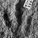
Les traces de pas de Dinosaures et autres Archosaures du Lias inférieur des grands Causses, Sud de la FranceGeorges Demathieu, Georges Gand, Jacques Sciau, Pierre Freytet and Jacques GarricPublished online: 15/12/2002Keywords: Dinosauroid footprints; France; Grands-Causses; Hettangian; ichnostratigraphy; paleoenvironments; Sinemurian; statistical results https://doi.org/10.18563/pv.31.1-4.1-143 Abstract The Causses" is a near 3400 km2 large plateau located in the south of France. Here the first dinosaur footprints where found in 1935. After this, this area has yielded an ever-increasing number of ichnites now in excess of 500 specimens. These latter, 15 to 50 cm long, tridactyl or tetradactyl footprints of generally biped animals, were discovered at the surface of Hettangian to lower Sinemurian dolomite layers within 4 distinct stratigraphic units. The 35 sites bearing ichnites are located on the plateau margin. For the first time, morphologic characters studied through descriptive statistic methods with the usual parameters and classical Student and Snédecor tests, allowed us, to divide the whole set of biped traces into 6 ichnospecies. Their definitions are further constrained by multivariate statistical results using Principal Component Analysis (PCA), Factor Analysis of correspondances (FAC) and Discriminant Analysis (DA). All have confirmed the morphologic observations. So that now, the following taxa are identified : Grallator variabilis, G. lescurei, G. sauclierensis, G. minusculus, Eubrontes giganteus, Dilophosauripus williamsi, cf. Moraesichnium, Orníthopus fabrei nov ichnosp. The more immediately visible differences relate to the interdigital II-IV divarication and the digit length ratio. To this panel, we must add Batrachopus deweyi and shapes suggesting Trisauropodichnus and/or Anomoepus. Among all ichnite associations described in the lower Liasic, the New England assemblage presents the most affinities with ours. It shows the ichnotaxa Grallator, Dilophosauripus, Eubrontes, Batrachopus without forgetting Ornithopus fabrei nov. ichnosp. which is close to Ornithopus gallinaceus from the Massachusetts and Connecticut basins. On comparing the present early Jurassic ichnofauna of the Causses with the ones of the Middle and Upper Triassic formations of the eastem border of the Massif Central (France), it appears that tridactyl footprints become more and more numerous and large from Triassic to Early Jurassic. In the Causses, these latest are prevalent but in Quercy (France), Poland, Italy, USA, they are also associated with Omithopoda, Thyreophora and Sauropoda ichnites. Footprint areas considered here were generaly under an arid climate. Animals that passed by were heavy and bulky possible Megalosaur trackmakers, and lighter and slender Coelophysids or Ceratosaurs. For all, these areas were pathways as the orientations of the trackways seem point out. The directions followed by these reptiles were without any important variation during the Hettango-Sinemurian stages. These areas were also used from time to time by Crocodilomorpha and may be tetradactyl (I-IV) bipedal avian Theropods. However, the number of such trackways in sites, sometimes substantial, should not lead us to overestimate the trackmakers populations. These last were probably relatively moderately abondant in this inter-supratidal swamp environment. In the Causses, ichnites are connected with former algo-laminated deposits (Algal mats) which were rapidly hardened by means of calcitisation of cyanobacteria. The result has been a moderate depth of footprints; autopodia disturbing only a few cm of the carbonate substrate. Other fossils have been discovered : invertebrates with thin bivalve and gastropod shells, crustaceans tests and plants. These latter suggest the existence of paleomangroves like environments but also continental vegetation periodically overruning the swamp environment during regression/transgression cycles. At these times, wooded parts of it, could become protecting, feeding, resting and nesting places. PV article infos Published in Vol. 31, Fasc. 1-4 (2002) |
|

|
Cervus elaphus rossii (Mammalia, Artiodactyla), a new endemic sub-species from the Middle Pleistocene of CorsicaElisabeth PereiraPublished online: 28/12/2001Keywords: Cervus elaphus; Corsica; Endemism; Pleistocene Abstract Several endemic deer remains from the Middle Pleistocene deposits of the Castiglione cave (Oletta, Haute-Corse) are examined here. A morphometric analysis allows to relate them to a new insular subspecies Cervus elaphus rossii. The bones were compared with those of the mainland early Middle Pleistocene subspecies Cervus elaphus acoronatus Beninde and the European species Cervus elaphus Linné (Late Middle Pleistocene and Upper Pleistocene forms (continental and insular)). The Castiglione fossil shows peculiar morphofunctional features in its appendicular skeleton suggesting a morphological convergence with certain Bovidae. PV article infos Published in Vol. 30, Fasc. 3-4 (2001) |
|

|
Cryptomerix Schlosser, 1886, Tragulidé de l'oligocène d'Europe ; relations du genre et considérations sur l'origine des ruminants.Jean SudrePublished online: 01/06/1984Keywords: Archaic Ruminants; Paleobiogeography; Quercy Phosphorites; Systematics; Tragulids https://doi.org/10.18563/pv.14.1.1-31 Abstract The genus Cryptomeryx SCHLOSSER, 1886, inusited during a long period, has been discovered in Lower and Middle Oligocene localities of the Quercy region (South-West France). This new material, as well as specimens from the old collections referred to Cryptomeryx, are described; their study, allows us precising the definition of the genus, and confirming its allocation to the Tragulidae family. The type species of the genus, Crypmmeryx gaudryi (= Lophiomeryx gaudryi FILHOL, 1877), occurs in several localities at the base of the Middle Oligocene (Itardies, La Plante 2, Roqueprune 2, Soulce, Herrlingen 1). The new species C. matsoui n. sp. has been defined in the older locality of Mas de Got (top of Lower Oligocene). It is possible that the species Pseudamphimeryx decedens STEHLIN, 1910 pertains to the same genus. Also to the Tragulids must be referred the monospecific genus Iberomeryx (I. parvus GABOUNIA, 1964) from Upper Oligocene of Benara (Georgie, URSS), with which Cryptomeryx is related. These genera are not direct ancestors of Miocene tragulids; their occurrence in the Western European Oligocene results from a first immigration wawe of the family. These Tragulids are one of the most archaic groups of Ruminants. They are probably derived from a primitive stock which had acquired in Asia the selenodont condition of the dentition. PV article infos Published in Vol. 14, Fasc. 1 (1984) |
|

|
Contributions à l'étude des micromammifères du gisement Miocène supérieur de Montredon (Hérault). 4 - Les chiroptèresBernard SigéPublished online: 30/06/1982Keywords: Chiroptera; Hérault; Late Miocene; Micromammals; Montredon https://doi.org/10.18563/pv.12.3.133-140 Abstract The Montredon local fauna yielded very rare bats, represented by damaged isolated teeth. Only a few documents are available for this period of the European Neogene. ln this poor state of knowledge, the material represents three undetermined species, a supposed molossid and two vespertilionids. PV article infos Published in Vol. 12, Fasc. 3 (1982) |
|

|
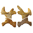
Etude de la Variabilité chez Lophiodon lautricense NouletJean SudrePublished online: 28/02/1971Keywords: Cheek teeth; Eocene; Lophiodon; variability https://doi.org/10.18563/pv.4.3.67-95 Abstract The biometric and morphologie variability of the cheek teeth in the end-of-the-phylum species Lophiodon lautricense Noulet studied in this note, reposes on the observation of about 800 teeth. These were revealed to be little variable in absolute dimensions. The considerable morphologie variability in the upper premolars permitted the problem of the molarization process to be taken up. An hypothesis concerning the order of eruption of the cheek teeth is formulated based on an examination of a large number of milk dentitions. In conclusion, it is suggested that reservations be held on the value of dental characters classically used in systematics for the group under consideration. PV article infos Published in Vol. 04, Fasc. 3 (1971) |
|

|
Le genre Plagiolophus (Palaeotheriidae, Perissodactyla, Mammalia): révision systématique, morphologie et histologie dentaires, anatomie crânienne, essai d'interprétation fonctionnelleJean-Albert RemyPublished online: 15/12/2004Keywords: New taxa; Paléogène; perissodactyls; skull anatomy; tooth histology Abstract The genus Plagiolophus is documented, almost solely in Western Europe, from the middle Eocene up to the mid Oligocene (MP 12 to MP 25), i.e. more than for 15 MY. Seventeen species are now recorded whose two of them are new, P. ringeadei nov. sp. and P. mamertensis nov. sp. Some anatomical variations and the deflection of certain evolutionary trends justify the distinction of three subgenera, Paloplotherium, Fraasiolophus nov. and Plagiolophus s.s. The genus displays a wide range in size and weight (between 10 and 150 kg). The detailed description of the skull of several species is here given for the first time. PV article infos Published in Vol. 33, Fasc. 1-4 (2004) |
|

|
The Ctenodactylidae (Rodentia) from the Oligocene of Ulantatal (inner Mongolia, China)Monique Vianey-Liaud
Published online: 15/12/2006 |
|
|
Nouvelles données sur les Ichnites de dinosaures d'El Bayadh (Crétacé Inférieur, Algérie)Mostefa Bessedik
Published online: 16/12/2008 |
|

|
Un gisement à mammifères dans la formation lacustre d'âge Miocène moyen du Collet Redon près de St-Cannat (Bouches-du-Rhone). Implications stratigaphiquesJean-Pierre Aguilar and G. ClauzonPublished online: 10/04/1979Keywords: France; Neogene; Rodentia https://doi.org/10.18563/pv.8.5.327-341 Abstract The new fauna of Collet Redon (Bouches-du-Rhône, France) is dated by three rodents: Megacricetodon aff. bavaricus, Democricetodon affinis mutilus and Peridyromys cf. hamadryas. They correlate this locality with Oggenhof and Ohningen in Bavaria (Western Germany). As the radiometric age of Ohningen is estimated between 14 and 13 M.Y., these three localities are of Serravallian age. This datation brings a complete readjusment of the stratigraphy of the section of Collet Redon formerly described by Collot and Combaluzier. The marine deposits with underly the continental formation with the mammal fauna, are Burdigalian. The angular unconformity between the marine and the continental deposits gives evidence of an episode of emersion on the margin of a sedimentary basin, with deformation and erosion. Owing to the newly discovered fauna, this geodynamical event is clearly settled within the regional geographical and chronological context. Lacustrine and continental deposits of such an age were up to now unsuspected in this area. PV article infos Published in Vol. 08, Fasc. 5 (1979) |
|

|
Les rongeurs du Miocène moyen et supérieur du MaghrebJean-Jacques Jaeger
Published online: 15/05/1977 |
|

|
Paleobiology of Messel ErinaceomorphsGerhard StorchPublished online: 16/12/1996Keywords: Erinaceomorpha; Germany; Grube Messel; Lipotyphla; Middle Eocene; Paleobiology Abstract Three erinaceomorph species are known from the early Middle Eocene of Grube Messel near Darmstadt, Germany, which are referred to the family Amphilemuridae. Pholidocercus hassiacus, Macrocranion tupaiodon, and Macrocranion tenerum showed extraordinary adaptations to their different life strategies, and several of their specializations are unknown among living insectivores. Pholídocercus was a well-defended robust animal with an opportunistic feeding strategy. Macrocraníon zupaiodon was a slender forest floor-dweller with saltatorial specializations to escape from predators; fishes were the preferred component of its omnivorous diet. Macrocranion tenerum exhibited a combination of both survival strategies, extremely elongated hind limbs for rapid and even ricochetal flight and a spiny exterior as an effective protective device; it was probably specialized for feeding on ants. Thus, closely related, omnivorous-insectivorous forest floor-dwellers could exploit the Messel ecosystem. PV article infos Published in Vol. 25, Fasc. 2-4 (1996) |
|

|
The Late Cretaceous nesting site of Auca Mahuevo (Patagonia, Argentina): eggs, nests, and embryos of titanosaurian sauropods.Luis M. Chiappe
Published online: 15/12/2003 |
|

|
Les Gruiformes (Aves) des phosphorites du Quercy (France). 1. sous-ordre cariamae (Cariamidae et Phorusrhacidae), systématique et biostratigraphie.Cécile Mourer-Chauviré
Published online: 30/11/1983 |
|
|
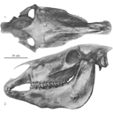
Old world hemiones and new world slender species (Mammalia, Equidae)Véra Eisenmann, John Howe and Mario PichardoPublished online: 16/12/2008Keywords: Amerhippus; biometry; Equus; Holocene; New World; Old World; Osteology; Pleistocene; Pliocene https://doi.org/10.18563/pv.36.1-4.159-233 Abstract Morphological and biometrical description of skulls, teeth, and limb bones of extant and fossil Old World herniones (including E. hydruntinus) and of New World 'stilt-Iegged' and other slender species from Blancan to Holocene. An Appendix presents ways in which the approximate size of some missing bones or dimensions may be deduced from available ones. PV article infos Published in Vol. 36, Fasc. 1-4 (2008) |
|

|
Eggshell microstructure and porosity of the Nicobar scrubfowl (Megapodius Nicobariensis, great Nicobar island, India)Géraldine Garcia
Published online: 16/12/2008 |
|

|
Book of Abstracts of the 20th Annual Conference of the European Association of Vertebrate Palaeontologists, 26th June – 1st July 2023, Sabadell (Barcelona), SpainDavid M. Alba
Published online: 15/06/2023 |
|
|
Latest Early-early Middle Eocene deposits of Algeria (Glib Zegdou, HGL50), yield the richest and most diverse fauna of amphibians and squamate reptiles from the Palaeogene of AfricaJean-Claude Rage
Published online: 08/02/2021 |
|
|
New material of “Eurysternidae” (Thalassochelydia, Pan-Cryptodira) from the Kimmeridgian of the Swiss Jura MountainsChristian Püntener
Published online: 25/06/2020 |
|